 Movie
Movie Controller

[English] 日本語
 Yorodumi
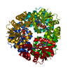
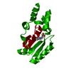
- PDB-3nd6: Crystal structure of phosphopantetheine adenylyltransferase (PPAT... -
+ Open data

- Basic information
| Entry | Database: PDB / ID: 3nd6 | ||||||
|---|---|---|---|---|---|---|---|
| Title | Crystal structure of phosphopantetheine adenylyltransferase (PPAT) in complex with ATP from Enterococcus faecalis | ||||||
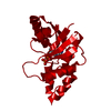
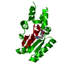
 Components Components | Phosphopantetheine adenylyltransferase | ||||||
 Keywords Keywords | TRANSFERASE / Phosphopantetheine adenylyltransferase / PPAT / Coenzyme A biosynthetic pathway | ||||||
| Function / homology |  Function and homology information Function and homology informationpantetheine-phosphate adenylyltransferase / pantetheine-phosphate adenylyltransferase activity / coenzyme A biosynthetic process / ATP binding / cytoplasm Similarity search - Function | ||||||
| Biological species |   Enterococcus faecalis (bacteria) Enterococcus faecalis (bacteria) | ||||||
| Method |  X-RAY DIFFRACTION / SYNCHROTRON / MOLECULAR REPLACEMENT / Resolution: 2.3 Å X-RAY DIFFRACTION / SYNCHROTRON / MOLECULAR REPLACEMENT / Resolution: 2.3 Å | ||||||
 Authors Authors | Yoon, H.J. / Lee, H.H. / Suh, S.W. | ||||||
 Citation Citation | Journal: Mol.Cells / Year: 2011 Title: Crystal structure of phosphopantetheine adenylyltransferase from Enterococcus faecalis in the ligand-unbound state and in complex with ATP and pantetheine Authors: Yoon, H.J. / Kang, J.Y. / Mikami, B. / Lee, H.H. / Suh, S.W. | ||||||
| History |
|
- Structure visualization
| Structure viewer | Molecule: MolmilJmol/JSmol |
|---|
- Downloads & links
-Download
| PDBx/mmCIF format | 3nd6.cif.gz | 205.9 KB | Display | PDBx/mmCIF format |
|---|---|---|---|---|
| PDB format | pdb3nd6.ent.gz | 164.7 KB | Display | PDB format |
| PDBx/mmJSON format | 3nd6.json.gz | Tree view | PDBx/mmJSON format | |
| Others |  Other downloads Other downloads |
-Validation report
| Arichive directory | https://data.pdbj.org/pub/pdb/validation_reports/nd/3nd6ftp://data.pdbj.org/pub/pdb/validation_reports/nd/3nd6 | HTTPS FTP |
|---|
-Related structure data
| Related structure data |  3nd5C  3nd7C 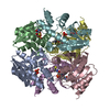 1vlhS C: citing same article ( S: Starting model for refinement |
|---|---|
| Similar structure data |


-Links
 PDBj
PDBj- Assembly
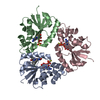
| Deposited unit | 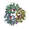
| ||||||||
|---|---|---|---|---|---|---|---|---|---|
| 1 |
| ||||||||
| Unit cell |
|
-Components
| #1: Protein | Mass: 19770.629 Da / Num. of mol.: 6 Source method: isolated from a genetically manipulated source Source: (gene. exp.) Enterococcus faecalis (bacteria) / Gene: coaD / Plasmid: pET-21a(+) / Production host: References: UniProt: Q831P9, pantetheine-phosphate adenylyltransferase #2: Chemical | ChemComp-ATP /   Mass: 507.181 Da / Num. of mol.: 6 / Source method: obtained synthetically / Formula: C10H16N5O13P3 / Comment: ATP, energy-carrying molecule*YM Mass: 507.181 Da / Num. of mol.: 6 / Source method: obtained synthetically / Formula: C10H16N5O13P3 / Comment: ATP, energy-carrying molecule*YM#3: Water | ChemComp-HOH / |  Mass: 18.015 Da / Num. of mol.: 413 / Source method: isolated from a natural source / Formula: H2O Mass: 18.015 Da / Num. of mol.: 413 / Source method: isolated from a natural source / Formula: H2O |
|---|
-Experimental details
-Experiment
| Experiment | Method: X-RAY DIFFRACTION / Number of used crystals: 1 |
|---|
- Sample preparation
| Crystal | Density Matthews: 3.66 Å3/Da / Density % sol: 66.39 % |
|---|---|
| Crystal grow | Temperature: 297 K / Method: vapor diffusion, hanging drop / pH: 8.5 Details: 3.5M sodium formate, 100mM Tris-HCl, pH 8.5, VAPOR DIFFUSION, HANGING DROP, temperature 297K |
-Data collection
| Diffraction | Mean temperature: 100 K |
|---|---|
| Diffraction source | Source: SYNCHROTRON / Site: Photon Factory  / Beamline: BL-17A / Wavelength: 1 Å / Beamline: BL-17A / Wavelength: 1 Å |
| Detector | Type: ADSC QUANTUM 270 / Detector: CCD / Date: Dec 22, 2006 / Details: Mirrors |
| Radiation | Monochromator: Numerical link type Si(111) / Protocol: SINGLE WAVELENGTH / Monochromatic (M) / Laue (L): M / Scattering type: x-ray |
| Radiation wavelength | Wavelength: 1 Å / Relative weight: 1 |
| Reflection | Resolution: 2.3→20 Å / Num. obs: 76960 / % possible obs: 98.9 % / Rmerge(I) obs: 0.065 / Net I/σ(I): 17.7 / Num. measured all: 534418 |
| Reflection shell | Resolution: 2.3→2.38 Å / Rmerge(I) obs: 0.232 / Mean I/σ(I) obs: 10.9 / Num. unique all: 7489 / % possible all: 97.9 |
- Processing
| Software |
| ||||||||||||||||||||
|---|---|---|---|---|---|---|---|---|---|---|---|---|---|---|---|---|---|---|---|---|---|
| Refinement | Method to determine structure: MOLECULAR REPLACEMENT Starting model: PDB ENTRY 1VLH Resolution: 2.3→20 Å / Cross valid method: THROUGHOUT / σ(F): 0
| ||||||||||||||||||||
| Displacement parameters | Biso mean: 44.2 Å2 | ||||||||||||||||||||
| Refinement step | Cycle: LAST / Resolution: 2.3→20 Å
|
