 Movie
Movie Controller

[English] 日本語
 Yorodumi
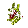
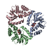
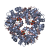
- PDB-6qmg: Phosphopantetheine adenylyltransferase from Mycobacterium tubercu... -
+ Open data

- Basic information
| Entry | Database: PDB / ID: 6qmg | |||||||||
|---|---|---|---|---|---|---|---|---|---|---|
| Title | Phosphopantetheine adenylyltransferase from Mycobacterium tuberculosis in complex with 5-methyl-1-phenyl-1H-pyrazole-4-carboxylic acid at 1.8A resolution. | |||||||||
 Components Components | Phosphopantetheine adenylyltransferase | |||||||||
 Keywords Keywords | TRANSFERASE / CoaD / PPAT / Complex | |||||||||
| Function / homology |  Function and homology information Function and homology informationpantetheine-phosphate adenylyltransferase / pantetheine-phosphate adenylyltransferase activity / coenzyme A biosynthetic process / protein hexamerization / ATP binding / cytoplasm Similarity search - Function | |||||||||
| Biological species |   Mycobacterium tuberculosis (bacteria) Mycobacterium tuberculosis (bacteria) | |||||||||
| Method |  X-RAY DIFFRACTION / SYNCHROTRON / MOLECULAR REPLACEMENT / Resolution: 1.65 Å X-RAY DIFFRACTION / SYNCHROTRON / MOLECULAR REPLACEMENT / Resolution: 1.65 Å | |||||||||
 Authors Authors | Blaszczyk, M. / Blundell, T.L. | |||||||||
| Funding support |  United States, 1items United States, 1items
| |||||||||
 Citation Citation | Journal: To Be Published Title: Fragment linking applied to the discovery of Mycobacterium tuberculosis phosphopantetheine adenylyltransferase inhibitors Authors: Blaszczyk, M. / El Bakali, J. / Boland, J.A. / Spry, C. / Dias, M. / Blundell, T.L. / Abel, C. | |||||||||
| History |
|
- Structure visualization
| Structure viewer | Molecule: MolmilJmol/JSmol |
|---|
- Downloads & links
-Download
| PDBx/mmCIF format | 6qmg.cif.gz | 45.4 KB | Display | PDBx/mmCIF format |
|---|---|---|---|---|
| PDB format | pdb6qmg.ent.gz | 31 KB | Display | PDB format |
| PDBx/mmJSON format | 6qmg.json.gz | Tree view | PDBx/mmJSON format | |
| Others |  Other downloads Other downloads |
-Validation report
| Arichive directory | https://data.pdbj.org/pub/pdb/validation_reports/qm/6qmgftp://data.pdbj.org/pub/pdb/validation_reports/qm/6qmg | HTTPS FTP |
|---|
-Related structure data
| Related structure data |  6g6vC  6qmfC  6qmhC  6qmiC  1tfuS S: Starting model for refinement C: citing same article ( |
|---|---|
| Similar structure data |


-Links
 PDBj
PDBj- Assembly
| Deposited unit | 
| |||||||||
|---|---|---|---|---|---|---|---|---|---|---|
| 1 | x 6
| |||||||||
| Unit cell |
| |||||||||
| Components on special symmetry positions |
|
-Components
| #1: Protein | Mass: 17792.385 Da / Num. of mol.: 1 Source method: isolated from a genetically manipulated source Source: (gene. exp.) Mycobacterium tuberculosis (strain ATCC 25618 / H37Rv) (bacteria)Strain: ATCC 25618 / H37Rv / Gene: coaD, kdtB, Rv2965c, MTCY349.22, u0002e / Production host: References: UniProt: P9WPA5, pantetheine-phosphate adenylyltransferase |
|---|---|
| #2: Chemical | ChemComp-IOP /   Mass: 189.211 Da / Num. of mol.: 1 / Source method: obtained synthetically / Formula: C11H11NO2 Mass: 189.211 Da / Num. of mol.: 1 / Source method: obtained synthetically / Formula: C11H11NO2 |
| #3: Water | ChemComp-HOH /  Mass: 18.015 Da / Num. of mol.: 73 / Source method: isolated from a natural source / Formula: H2O Mass: 18.015 Da / Num. of mol.: 73 / Source method: isolated from a natural source / Formula: H2O |
-Experimental details
-Experiment
| Experiment | Method: X-RAY DIFFRACTION / Number of used crystals: 1 |
|---|
- Sample preparation
| Crystal | Density Matthews: 2.94 Å3/Da / Density % sol: 58.2 % / Description: cubes |
|---|---|
| Crystal grow | Temperature: 293 K / Method: vapor diffusion, sitting drop / pH: 7.5 / Details: MPD, CoHexamine Chloride, Cacodylate/Tris / PH range: 6.5-8.5 |
-Data collection
| Diffraction | Mean temperature: 100 K / Serial crystal experiment: N | ||||||||||||||||||||||||||||||||||||||||||||||||||||||||||||||||||||||||||||||||||||||||
|---|---|---|---|---|---|---|---|---|---|---|---|---|---|---|---|---|---|---|---|---|---|---|---|---|---|---|---|---|---|---|---|---|---|---|---|---|---|---|---|---|---|---|---|---|---|---|---|---|---|---|---|---|---|---|---|---|---|---|---|---|---|---|---|---|---|---|---|---|---|---|---|---|---|---|---|---|---|---|---|---|---|---|---|---|---|---|---|---|---|
| Diffraction source | Source: SYNCHROTRON / Site: Diamond  / Beamline: I03 / Wavelength: 0.98 Å / Beamline: I03 / Wavelength: 0.98 Å | ||||||||||||||||||||||||||||||||||||||||||||||||||||||||||||||||||||||||||||||||||||||||
| Detector | Type: DECTRIS PILATUS3 6M / Detector: PIXEL / Date: Jul 2, 2012 | ||||||||||||||||||||||||||||||||||||||||||||||||||||||||||||||||||||||||||||||||||||||||
| Radiation | Protocol: SINGLE WAVELENGTH / Monochromatic (M) / Laue (L): M / Scattering type: x-ray | ||||||||||||||||||||||||||||||||||||||||||||||||||||||||||||||||||||||||||||||||||||||||
| Radiation wavelength | Wavelength: 0.98 Å / Relative weight: 1 | ||||||||||||||||||||||||||||||||||||||||||||||||||||||||||||||||||||||||||||||||||||||||
| Reflection | Resolution: 1.648→67.923 Å / Num. all: 25454 / Num. obs: 25454 / % possible obs: 100 % / Redundancy: 19.7 % / Biso Wilson estimate: 29.04 Å2 / Rpim(I) all: 0.016 / Rrim(I) all: 0.072 / Rsym value: 0.067 / Net I/av σ(I): 5.1 / Net I/σ(I): 26.4 / Num. measured all: 501122 | ||||||||||||||||||||||||||||||||||||||||||||||||||||||||||||||||||||||||||||||||||||||||
| Reflection shell | Diffraction-ID: 1 / % possible all: 100
|
- Processing
| Software |
| ||||||||||||||||||||||||||||||||||||||||||||||||||||||||||||||||||||||
|---|---|---|---|---|---|---|---|---|---|---|---|---|---|---|---|---|---|---|---|---|---|---|---|---|---|---|---|---|---|---|---|---|---|---|---|---|---|---|---|---|---|---|---|---|---|---|---|---|---|---|---|---|---|---|---|---|---|---|---|---|---|---|---|---|---|---|---|---|---|---|---|
| Refinement | Method to determine structure: MOLECULAR REPLACEMENT Starting model: 1TFU Resolution: 1.65→67.92 Å / SU ML: 0.24 / Cross valid method: THROUGHOUT / σ(F): 1.35 / Phase error: 26.06
| ||||||||||||||||||||||||||||||||||||||||||||||||||||||||||||||||||||||
| Solvent computation | Shrinkage radii: 0.9 Å / VDW probe radii: 1.11 Å | ||||||||||||||||||||||||||||||||||||||||||||||||||||||||||||||||||||||
| Displacement parameters | Biso mean: 35.5 Å2 | ||||||||||||||||||||||||||||||||||||||||||||||||||||||||||||||||||||||
| Refinement step | Cycle: LAST / Resolution: 1.65→67.92 Å
| ||||||||||||||||||||||||||||||||||||||||||||||||||||||||||||||||||||||
| Refine LS restraints |
| ||||||||||||||||||||||||||||||||||||||||||||||||||||||||||||||||||||||
| LS refinement shell |
|
