[English] 日本語
Yorodumi
- PDB-3nbd: Clitocybe nebularis ricin B-like lectin (CNL) in complex with lac... -

+
Open data


ID or keywords:

Loading...

-
Basic information

Entry
Database: PDB / ID: 3nbd
TitleClitocybe nebularis ricin B-like lectin (CNL) in complex with lactose, crystallized at pH 7.1
ComponentsRicin B-like lectin
KeywordsSUGAR BINDING PROTEIN / Clitocybe nebularis ricin B-like lectin / lactose
Function / homology
Function and homology information


carbohydrate binding / protein homodimerization activity
Similarity search - Function
Ricin-type beta-trefoil lectin domain-like / Ricin B, lectin domain / Ricin B-like lectins / Trefoil (Acidic Fibroblast Growth Factor, subunit A) - #50 / Trefoil (Acidic Fibroblast Growth Factor, subunit A) / Trefoil / Mainly Beta
Similarity search - Domain/homology
alpha-lactose / Ricin B-like lectin
Similarity search - Component
Biological speciesClitocybe nebularis (fungus)
MethodX-RAY DIFFRACTION / SYNCHROTRON / MOLECULAR REPLACEMENT / Resolution: 1.15 Å
AuthorsRenko, M. / Pohleven, J. / Sabotic, J. / Kos, J. / Turk, D.
Citation
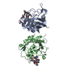
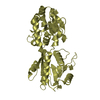
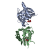
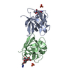
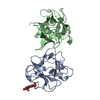
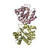
Journal: J.Biol.Chem. / Year: 2012
Title: Bivalent carbohydrate binding is required for biological activity of Clitocybe nebularis lectin (CNL), the N,N'-diacetyllactosediamine (GalNAc beta 1-4GlcNAc, LacdiNAc)-specific lectin from ...Title: Bivalent carbohydrate binding is required for biological activity of Clitocybe nebularis lectin (CNL), the N,N'-diacetyllactosediamine (GalNAc beta 1-4GlcNAc, LacdiNAc)-specific lectin from basidiomycete C. nebularis
Authors: Pohleven, J. / Renko, M. / Magister, S. / Smith, D.F. / Kunzler, M. / Strukelj, B. / Turk, D. / Kos, J. / Sabotic, J.
History
DepositionJun 3, 2010Deposition site: RCSB / Processing site: PDBJ
Revision 1.0Sep 21, 2011Provider: repository / Type: Initial release
Revision 1.1Mar 7, 2012Group: Database references
Revision 1.2Mar 19, 2014Group: Database references
Revision 1.3Nov 8, 2017Group: Advisory / Refinement description
Category: pdbx_unobs_or_zero_occ_atoms / pdbx_unobs_or_zero_occ_residues / software
Revision 2.0Jul 29, 2020Group: Atomic model / Data collection ...Atomic model / Data collection / Derived calculations / Non-polymer description / Structure summary
Category: atom_site / atom_site_anisotrop ...atom_site / atom_site_anisotrop / chem_comp / entity / entity_name_com / pdbx_branch_scheme / pdbx_chem_comp_identifier / pdbx_entity_branch / pdbx_entity_branch_descriptor / pdbx_entity_branch_link / pdbx_entity_branch_list / pdbx_entity_nonpoly / pdbx_molecule_features / pdbx_nonpoly_scheme / struct_asym / struct_conn / struct_site / struct_site_gen
Item: _atom_site.B_iso_or_equiv / _atom_site.Cartn_x ..._atom_site.B_iso_or_equiv / _atom_site.Cartn_x / _atom_site.Cartn_y / _atom_site.Cartn_z / _atom_site.auth_asym_id / _atom_site.auth_atom_id / _atom_site.auth_comp_id / _atom_site.auth_seq_id / _atom_site.label_asym_id / _atom_site.label_atom_id / _atom_site.label_comp_id / _atom_site.label_entity_id / _atom_site.type_symbol / _atom_site_anisotrop.U[1][1] / _atom_site_anisotrop.U[1][2] / _atom_site_anisotrop.U[1][3] / _atom_site_anisotrop.U[2][2] / _atom_site_anisotrop.U[2][3] / _atom_site_anisotrop.U[3][3] / _atom_site_anisotrop.pdbx_auth_asym_id / _atom_site_anisotrop.pdbx_auth_atom_id / _atom_site_anisotrop.pdbx_auth_comp_id / _atom_site_anisotrop.pdbx_auth_seq_id / _atom_site_anisotrop.pdbx_label_asym_id / _atom_site_anisotrop.pdbx_label_atom_id / _atom_site_anisotrop.pdbx_label_comp_id / _atom_site_anisotrop.type_symbol / _chem_comp.formula / _chem_comp.formula_weight / _chem_comp.id / _chem_comp.mon_nstd_flag / _chem_comp.name / _chem_comp.type / _entity.formula_weight / _entity.pdbx_description / _entity.type / _struct_asym.entity_id
Description: Carbohydrate remediation / Provider: repository / Type: Remediation
Revision 2.1Nov 1, 2023Group: Advisory / Data collection ...Advisory / Data collection / Database references / Refinement description / Structure summary
Category: chem_comp / chem_comp_atom ...chem_comp / chem_comp_atom / chem_comp_bond / database_2 / pdbx_initial_refinement_model / pdbx_unobs_or_zero_occ_atoms / pdbx_unobs_or_zero_occ_residues
Item: _chem_comp.pdbx_synonyms / _database_2.pdbx_DOI / _database_2.pdbx_database_accession

-
Structure visualization

Structure viewerMolecule:
MolmilJmol/JSmol

Downloads & links

-
Assembly

Deposited unit
A: Ricin B-like lectin
B: Ricin B-like lectin
hetero molecules


Theoretical massNumber of molelcules
Total (without water)32,9069
Polymers31,7412
Non-polymers1,1657
Water9,638535
1


  • Idetical with deposited unit
  • defined by author&software
TypeNameSymmetry operationNumber
identity operation1_555x,y,z1
Buried area1280 Å2
ΔGint-12 kcal/mol
Surface area13820 Å2
MethodPISA
Unit cell
Length a, b, c (Å)43.119, 85.080, 97.629
Angle α, β, γ (deg.)90.00, 90.00, 90.00
Int Tables number19
Space group name H-MP212121

-
Components

#1: Protein Ricin B-like lectin


Mass: 15870.543 Da / Num. of mol.: 2
Source method: isolated from a genetically manipulated source
Source: (gene. exp.) Clitocybe nebularis (fungus) / Plasmid: pET11a / Production host: Escherichia coli (E. coli) / Strain (production host): BL21(DE3) / References: UniProt: B2ZRS9
#2: Polysaccharide beta-D-galactopyranose-(1-4)-alpha-D-glucopyranose / alpha-lactose


Type: oligosaccharide, Oligosaccharide / Class: Nutrient / Mass: 342.297 Da / Num. of mol.: 2
Source method: isolated from a genetically manipulated source
Details: oligosaccharide / References: alpha-lactose
DescriptorTypeProgram
DGalpb1-4DGlcpa1-ROHGlycam Condensed SequenceGMML 1.0
WURCS=2.0/2,2,1/[a2122h-1a_1-5][a2112h-1b_1-5]/1-2/a4-b1WURCSPDB2Glycan 1.1.0
[][a-D-Glcp]{[(4+1)][b-D-Galp]{}}LINUCSPDB-CARE
#3: Chemical
ChemComp-SO4 / SULFATE ION


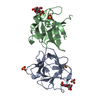
Mass: 96.063 Da / Num. of mol.: 5 / Source method: obtained synthetically / Formula: SO4
#4: Water ChemComp-HOH / water


Mass: 18.015 Da / Num. of mol.: 535 / Source method: isolated from a natural source / Formula: H2O

-
Experimental details

-
Experiment

ExperimentMethod: X-RAY DIFFRACTION / Number of used crystals: 1

-
Sample preparation

CrystalDensity Matthews: 2.82 Å3/Da / Density % sol: 56.4 % / Mosaicity: 0.43 °
Crystal growTemperature: 293 K / Method: vapor diffusion, sitting drop / pH: 7.1
Details: 0.085M HEPES sodium, 1.7%(v/v) PEG 400, 2.0M Ammonium sulfate, 19%(w/v) Glycerol, pH 7.1, VAPOR DIFFUSION, SITTING DROP, temperature 293K

-
Data collection

DiffractionMean temperature: 100 K
Diffraction sourceSource: SYNCHROTRON / Site: ELETTRA / Beamline: 5.2R / Wavelength: 1.033 Å
DetectorType: MAR CCD 165 mm / Detector: CCD / Date: Feb 18, 2009 / Details: Collimating and focusing, Pt-coated mirrors
RadiationMonochromator: Double Crystal Si111 / Protocol: SINGLE WAVELENGTH / Monochromatic (M) / Laue (L): M / Scattering type: x-ray
Radiation wavelengthWavelength: 1.033 Å / Relative weight: 1
ReflectionRedundancy: 8.5 % / Av σ(I) over netI: 48.22 / Number: 1086560 / Rmerge(I) obs: 0.06 / Χ2: 1.73 / D res high: 1.14 Å / D res low: 23 Å / Num. obs: 128514 / % possible obs: 98.3
Diffraction reflection shell
Highest resolution (Å)Lowest resolution (Å)% possible obs (%)IDRmerge(I) obsChi squaredRedundancy
3.092399.910.0291.6078.8
2.463.0910010.0411.8549.8
2.152.4610010.0662.61115.7
1.952.1510010.0752.48915.3
1.811.9510010.0912.17415.3
1.71.8110010.1092.1310
1.621.710010.1232.1467
1.551.6210010.1342.0056.9
1.491.5510010.1461.7596.9
1.441.4910010.1591.5076.9
1.391.4410010.1741.3566.9
1.351.3910010.1871.2726.8
1.321.3510010.2031.1626.8
1.281.3210010.2171.1186.7
1.251.2810010.2351.0476.7
1.231.2510010.2520.9966.7
1.21.2310010.2690.9356.6
1.181.210010.290.8136.5
1.161.1810010.3110.6616.3
1.141.1665.610.3260.5595
ReflectionResolution: 1.14→23.03 Å / Num. all: 130730 / Num. obs: 128514 / % possible obs: 98.3 % / Observed criterion σ(F): 1 / Observed criterion σ(I): 1 / Redundancy: 8.5 % / Rmerge(I) obs: 0.06 / Χ2: 1.726 / Net I/σ(I): 11.5
Reflection shellResolution: 1.14→1.16 Å / Redundancy: 5 % / Rmerge(I) obs: 0.326 / Num. unique all: 4260 / Χ2: 0.559 / % possible all: 65.6

-
Processing

Software
NameVersionClassificationNB
DENZOdata reduction
SCALEPACKdata scaling
REFMACrefinement
PDB_EXTRACT3.1data extraction
MAR345dtbdata collection
AMoREphasing
RefinementMethod to determine structure: MOLECULAR REPLACEMENT
Starting model: 3NBC
Resolution: 1.15→23.03 Å / Cor.coef. Fo:Fc: 0.977 / Cor.coef. Fo:Fc free: 0.97 / WRfactor Rfree: 0.156 / WRfactor Rwork: 0.135 / Occupancy max: 1 / Occupancy min: 0 / FOM work R set: 0.944 / SU B: 0.608 / SU ML: 0.013 / SU R Cruickshank DPI: 0.028 / SU Rfree: 0.029 / Cross valid method: THROUGHOUT / σ(F): 0 / ESU R Free: 0.029 / Stereochemistry target values: MAXIMUM LIKELIHOOD
Details: HYDROGENS HAVE BEEN ADDED IN THE RIDING POSITIONS U VALUES: REFINED INDIVIDUALLY. The structure was refined also with MAIN.
RfactorNum. reflection% reflectionSelection details
Rfree0.16 6458 5 %RANDOM
Rwork0.138 ---
all0.142 130730 --
obs0.139 128492 99.51 %-
Solvent computationIon probe radii: 0.8 Å / Shrinkage radii: 0.8 Å / VDW probe radii: 1.4 Å / Solvent model: MASK
Displacement parametersBiso max: 68.6 Å2 / Biso mean: 15.915 Å2 / Biso min: 3.8 Å2
Baniso -1Baniso -2Baniso -3
1-0.33 Å20 Å20 Å2
2---0.01 Å20 Å2
3----0.32 Å2
Refinement stepCycle: LAST / Resolution: 1.15→23.03 Å
ProteinNucleic acidLigandSolventTotal
Num. atoms2238 0 71 535 2844
Refine LS restraints
Refine-IDTypeDev idealDev ideal targetNumber
X-RAY DIFFRACTIONr_bond_refined_d0.050.0412441
X-RAY DIFFRACTIONr_bond_other_d0.0380.0391532
X-RAY DIFFRACTIONr_angle_refined_deg2.2311.9633391
X-RAY DIFFRACTIONr_angle_other_deg1.6892.9973837
X-RAY DIFFRACTIONr_dihedral_angle_1_deg8.5665331
X-RAY DIFFRACTIONr_dihedral_angle_2_deg35.05926.154104
X-RAY DIFFRACTIONr_dihedral_angle_3_deg10.97215374
X-RAY DIFFRACTIONr_dihedral_angle_4_deg19.342158
X-RAY DIFFRACTIONr_chiral_restr0.8160.2417
X-RAY DIFFRACTIONr_gen_planes_refined0.0130.0212713
X-RAY DIFFRACTIONr_gen_planes_other0.0020.02416
X-RAY DIFFRACTIONr_mcbond_it2.3611.51522
X-RAY DIFFRACTIONr_mcbond_other0.9461.5606
X-RAY DIFFRACTIONr_mcangle_it3.42722514
X-RAY DIFFRACTIONr_scbond_it4.8633919
X-RAY DIFFRACTIONr_scangle_it6.8294.5855
X-RAY DIFFRACTIONr_rigid_bond_restr2.22633973
LS refinement shellResolution: 1.147→1.177 Å / Total num. of bins used: 20
RfactorNum. reflection% reflection
Rfree0.374 443 -
Rwork0.362 8401 -
all-8844 -
obs--93.66 %

+
About Yorodumi

-
News

-
Feb 9, 2022. New format data for meta-information of EMDB entries

New format data for meta-information of EMDB entries

  • Version 3 of the EMDB header file is now the official format.
  • The previous official version 1.9 will be removed from the archive.

Related info.:EMDB header

External links:wwPDB to switch to version 3 of the EMDB data model

-
Aug 12, 2020. Covid-19 info

Covid-19 info

URL: https://pdbjlvh1.pdbj.org/emnavi/covid19.php

New page: Covid-19 featured information page in EM Navigator.

Related info.:Covid-19 info / Mar 5, 2020. Novel coronavirus structure data

+
Mar 5, 2020. Novel coronavirus structure data

Novel coronavirus structure data

Related info.:Yorodumi Speices / Aug 12, 2020. Covid-19 info

External links:COVID-19 featured content - PDBj / Molecule of the Month (242):Coronavirus Proteases

+
Jan 31, 2019. EMDB accession codes are about to change! (news from PDBe EMDB page)

EMDB accession codes are about to change! (news from PDBe EMDB page)

  • The allocation of 4 digits for EMDB accession codes will soon come to an end. Whilst these codes will remain in use, new EMDB accession codes will include an additional digit and will expand incrementally as the available range of codes is exhausted. The current 4-digit format prefixed with “EMD-” (i.e. EMD-XXXX) will advance to a 5-digit format (i.e. EMD-XXXXX), and so on. It is currently estimated that the 4-digit codes will be depleted around Spring 2019, at which point the 5-digit format will come into force.
  • The EM Navigator/Yorodumi systems omit the EMD- prefix.

Related info.:Q: What is EMD? / ID/Accession-code notation in Yorodumi/EM Navigator

External links:EMDB Accession Codes are Changing Soon! / Contact to PDBj

+
Jul 12, 2017. Major update of PDB

Major update of PDB

  • wwPDB released updated PDB data conforming to the new PDBx/mmCIF dictionary.
  • This is a major update changing the version number from 4 to 5, and with Remediation, in which all the entries are updated.
  • In this update, many items about electron microscopy experimental information are reorganized (e.g. em_software).
  • Now, EM Navigator and Yorodumi are based on the updated data.

External links:wwPDB Remediation / Enriched Model Files Conforming to OneDep Data Standards Now Available in the PDB FTP Archive

-
Yorodumi

Thousand views of thousand structures

  • Yorodumi is a browser for structure data from EMDB, PDB, SASBDB, etc.
  • This page is also the successor to EM Navigator detail page, and also detail information page/front-end page for Omokage search.
  • The word "yorodu" (or yorozu) is an old Japanese word meaning "ten thousand". "mi" (miru) is to see.

Related info.:EMDB / PDB / SASBDB / Comparison of 3 databanks / Yorodumi Search / Aug 31, 2016. New EM Navigator & Yorodumi / Yorodumi Papers / Jmol/JSmol / Function and homology information / Changes in new EM Navigator and Yorodumi

Read more