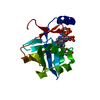
RNA exonuclease activity / DNA ligase (ATP) / DNA ligase (ATP) activity / DNA replication, synthesis of primer / DNA biosynthetic process / nucleotidyltransferase activity / double-strand break repair via nonhomologous end joining / DNA recombination / DNA-directed DNA polymerase activity / DNA binding ...RNA exonuclease activity / DNA ligase (ATP) / DNA ligase (ATP) activity / DNA replication, synthesis of primer / DNA biosynthetic process / nucleotidyltransferase activity / double-strand break repair via nonhomologous end joining / DNA recombination / DNA-directed DNA polymerase activity / DNA binding / ATP binding / metal ion binding Similarity search - Function
DNA ligase D / LigD polymerase domain, PaeLigD-type / DNA ligase D, ligase domain / DNA ligase D, polymerase domain / : / LigD, primase-polymerase domain / DNA ligase D, 3'-phosphoesterase domain / DNA Ligase D 3'-phosphoesterase domain / DNA ligase, ATP-dependent, C-terminal / ATP dependent DNA ligase C terminal region ...DNA ligase D / LigD polymerase domain, PaeLigD-type / DNA ligase D, ligase domain / DNA ligase D, polymerase domain / : / LigD, primase-polymerase domain / DNA ligase D, 3'-phosphoesterase domain / DNA Ligase D 3'-phosphoesterase domain / DNA ligase, ATP-dependent, C-terminal / ATP dependent DNA ligase C terminal region / ATP-dependent DNA ligase family profile. / DNA ligase, ATP-dependent, central / ATP dependent DNA ligase domain / Nucleic acid-binding, OB-fold Similarity search - Domain/homology
Mass: 18.015 Da / Num. of mol.: 85 / Source method: isolated from a natural source / Formula: H2O
-
Experimental details
-
Experiment
Experiment
Method: X-RAY DIFFRACTION / Number of used crystals: 1
-
Sample preparation
Crystal
Density Matthews: 2.53 Å3/Da / Density % sol: 51.4 %
Crystal grow
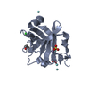
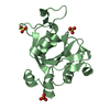
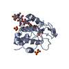
Temperature: 298 K / Method: vapor diffusion, sitting drop / pH: 6.5 Details: ;Crystallization was carried out in sitting-drop vapor-diffusion setups with 1:1 mixtures of protein solution containing 1.3 mM PaePEC2 and 2mM MnCl2 and reservoir solution containing PEG ...Details: ;Crystallization was carried out in sitting-drop vapor-diffusion setups with 1:1 mixtures of protein solution containing 1.3 mM PaePEC2 and 2mM MnCl2 and reservoir solution containing PEG 5000 monomethylether (MME) (20 - 30%), 100 mM 2-(N-morpholino) ethanesulfonic acid (MES) pH 6.8 - 7.0, 200 mM ammonium sulfate, and 10 mM yttrium (III) chloride at 22 C. , VAPOR DIFFUSION, SITTING DROP, temperature 298K
In the structure databanks used in Yorodumi, some data are registered as the other names, "COVID-19 virus" and "2019-nCoV". Here are the details of the virus and the list of structure data.
Jan 31, 2019. EMDB accession codes are about to change! (news from PDBe EMDB page)
EMDB accession codes are about to change! (news from PDBe EMDB page)
The allocation of 4 digits for EMDB accession codes will soon come to an end. Whilst these codes will remain in use, new EMDB accession codes will include an additional digit and will expand incrementally as the available range of codes is exhausted. The current 4-digit format prefixed with “EMD-” (i.e. EMD-XXXX) will advance to a 5-digit format (i.e. EMD-XXXXX), and so on. It is currently estimated that the 4-digit codes will be depleted around Spring 2019, at which point the 5-digit format will come into force.
The EM Navigator/Yorodumi systems omit the EMD- prefix.
Related info.:Q: What is EMD? / ID/Accession-code notation in Yorodumi/EM Navigator
Yorodumi is a browser for structure data from EMDB, PDB, SASBDB, etc.
This page is also the successor to EM Navigator detail page, and also detail information page/front-end page for Omokage search.
The word "yorodu" (or yorozu) is an old Japanese word meaning "ten thousand". "mi" (miru) is to see.
Related info.:EMDB / PDB / SASBDB / Comparison of 3 databanks / Yorodumi Search / Aug 31, 2016. New EM Navigator & Yorodumi / Yorodumi Papers / Jmol/JSmol / Function and homology information / Changes in new EM Navigator and Yorodumi
 Movie
Movie Controller

 Yorodumi
 Open data
 Basic information
 Components
 Keywords
 Function and homology information
 Pseudomonas aeruginosa (bacteria)
 X-RAY DIFFRACTION /
  Authors
 Citation
 Structure visualization
 Downloads & links
 Other downloads


 PDBj
PDBj


 Assembly
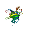

 Mass: 54.938 Da / Num. of mol.: 1 / Source method: obtained synthetically / Formula: Mn
 Mass: 96.063 Da / Num. of mol.: 2 / Source method: obtained synthetically / Formula: SO4
 Mass: 88.906 Da / Num. of mol.: 2 / Source method: obtained synthetically / Formula: Y
 Mass: 18.015 Da / Num. of mol.: 85 / Source method: isolated from a natural source / Formula: H2O
 Sample preparation
 Processing