 Movie
Movie Controller

[English] 日本語
 Yorodumi
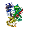
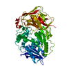
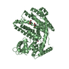
- PDB-3n98: Crystal structure of TK1436, a GH57 branching enzyme from hyperth... -
+ Open data

- Basic information
| Entry | Database: PDB / ID: 3n98 | ||||||
|---|---|---|---|---|---|---|---|
| Title | Crystal structure of TK1436, a GH57 branching enzyme from hyperthermophilic archaeon Thermococcus kodakaraensis, in complex with glucose and additives | ||||||
 Components Components | alpha-amylase, GH57 family | ||||||
 Keywords Keywords | TRANSFERASE / GH57 family member / branching enzyme | ||||||
| Function / homology |  Function and homology information Function and homology informationalpha-glucan biosynthetic process / 1,4-alpha-glucan branching enzyme / 1,4-alpha-glucan branching enzyme activity / nucleotide binding Similarity search - Function | ||||||
| Biological species |   Thermococcus kodakarensis (archaea) Thermococcus kodakarensis (archaea) | ||||||
| Method |  X-RAY DIFFRACTION / SYNCHROTRON / MOLECULAR REPLACEMENT / Resolution: 1.87 Å X-RAY DIFFRACTION / SYNCHROTRON / MOLECULAR REPLACEMENT / Resolution: 1.87 Å | ||||||
 Authors Authors | Santos, C.R. / Tonoli, C.C.C. / Trindade, D.M. / Betzel, C. / Takata, H. / Kuriki, T. / Kanai, T. / Imanaka, T. / Arni, R.K. / Murakami, M.T. | ||||||
 Citation Citation | Journal: Proteins / Year: 2011 Title: Structural basis for branching-enzyme activity of glycoside hydrolase family 57: Structure and stability studies of a novel branching enzyme from the hyperthermophilic archaeon Thermococcus Kodakaraensis KOD1. Authors: Santos, C.R. / Tonoli, C.C. / Trindade, D.M. / Betzel, C. / Takata, H. / Kuriki, T. / Kanai, T. / Imanaka, T. / Arni, R.K. / Murakami, M.T. | ||||||
| History |
|
- Structure visualization
| Structure viewer | Molecule: MolmilJmol/JSmol |
|---|
- Downloads & links
-Download
| PDBx/mmCIF format | 3n98.cif.gz | 140.2 KB | Display | PDBx/mmCIF format |
|---|---|---|---|---|
| PDB format | pdb3n98.ent.gz | 107.7 KB | Display | PDB format |
| PDBx/mmJSON format | 3n98.json.gz | Tree view | PDBx/mmJSON format | |
| Others |  Other downloads Other downloads |
-Validation report
| Arichive directory | https://data.pdbj.org/pub/pdb/validation_reports/n9/3n98ftp://data.pdbj.org/pub/pdb/validation_reports/n9/3n98 | HTTPS FTP |
|---|
-Related structure data
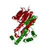
| Related structure data |  3n8tC  3n92C  1ufaS C: citing same article ( S: Starting model for refinement |
|---|---|
| Similar structure data |


-Links
 PDBj
PDBj


- Assembly
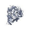
| Deposited unit | 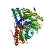
| ||||||||
|---|---|---|---|---|---|---|---|---|---|
| 1 |
| ||||||||
| Unit cell |
|
-Components
-Protein / Sugars , 2 types, 4 molecules A
| #1: Protein | Mass: 66167.953 Da / Num. of mol.: 1 / Fragment: UNP residues 1-562 Source method: isolated from a genetically manipulated source Source: (gene. exp.) Thermococcus kodakarensis (archaea) / Strain: KOD1 / Gene: TK1436 / Plasmid: pET21a(+) / Production host:  References: UniProt: Q5JDJ7, 1,4-alpha-glucan branching enzyme |
|---|---|
| #2: Sugar |  Type: D-saccharide, beta linking / Mass: 180.156 Da / Num. of mol.: 3 Type: D-saccharide, beta linking / Mass: 180.156 Da / Num. of mol.: 3Source method: isolated from a genetically manipulated source Formula: C6H12O6 |
-Non-polymers , 5 types, 355 molecules 


| #3: Chemical |  Mass: 92.094 Da / Num. of mol.: 3 / Source method: obtained synthetically / Formula: C3H8O3 Mass: 92.094 Da / Num. of mol.: 3 / Source method: obtained synthetically / Formula: C3H8O3#4: Chemical |  Mass: 88.105 Da / Num. of mol.: 2 / Source method: obtained synthetically / Formula: C4H8O2 Mass: 88.105 Da / Num. of mol.: 2 / Source method: obtained synthetically / Formula: C4H8O2#5: Chemical | ChemComp-PEG / |  Mass: 106.120 Da / Num. of mol.: 1 / Source method: obtained synthetically / Formula: C4H10O3 Mass: 106.120 Da / Num. of mol.: 1 / Source method: obtained synthetically / Formula: C4H10O3#6: Chemical | ChemComp-PG4 / |  Mass: 194.226 Da / Num. of mol.: 1 / Source method: obtained synthetically / Formula: C8H18O5 / Comment: precipitant*YM Mass: 194.226 Da / Num. of mol.: 1 / Source method: obtained synthetically / Formula: C8H18O5 / Comment: precipitant*YM#7: Water | ChemComp-HOH / | Mass: 18.015 Da / Num. of mol.: 348 / Source method: isolated from a natural source / Formula: H2O |
|---|
-Experimental details
-Experiment
| Experiment | Method: X-RAY DIFFRACTION / Number of used crystals: 1 |
|---|
- Sample preparation
| Crystal | Density Matthews: 2.83 Å3/Da / Density % sol: 56.53 % |
|---|---|
| Crystal grow | Temperature: 291 K / Method: vapor diffusion, sitting drop / pH: 5.5 Details: 100 mM sodium acetate (pH 5.5), 15% (w/v) PEG 8000, 10% (v/v) PEG 400, 200 mM sodium chloride, 1% (v/v) dioxane and 5% (v/v) glycerol , VAPOR DIFFUSION, SITTING DROP, temperature 291K |
-Data collection
| Diffraction | Mean temperature: 100 K |
|---|---|
| Diffraction source | Source: SYNCHROTRON / Site: LNLS  / Beamline: W01B-MX2 / Wavelength: 1.4586 Å / Beamline: W01B-MX2 / Wavelength: 1.4586 Å |
| Detector | Type: MARMOSAIC 225 mm CCD / Detector: CCD / Date: Feb 10, 2010 |
| Radiation | Monochromator: Si(111) double-crystal / Protocol: SINGLE WAVELENGTH / Monochromatic (M) / Laue (L): M / Scattering type: x-ray |
| Radiation wavelength | Wavelength: 1.4586 Å / Relative weight: 1 |
| Reflection | Resolution: 1.87→68.08 Å / Num. obs: 59969 / % possible obs: 95.4 % / Redundancy: 10.8 % / Biso Wilson estimate: 24.6 Å2 / Rmerge(I) obs: 0.104 / Net I/σ(I): 24 |
| Reflection shell | Resolution: 1.87→1.94 Å / Redundancy: 4.8 % / Rmerge(I) obs: 0.481 / Mean I/σ(I) obs: 2.2 / Num. unique all: 5216 / % possible all: 84.3 |
- Processing
| Software |
| |||||||||||||||||||||||||||||||||||||||||||||||||||||||||||||||||
|---|---|---|---|---|---|---|---|---|---|---|---|---|---|---|---|---|---|---|---|---|---|---|---|---|---|---|---|---|---|---|---|---|---|---|---|---|---|---|---|---|---|---|---|---|---|---|---|---|---|---|---|---|---|---|---|---|---|---|---|---|---|---|---|---|---|---|
| Refinement | Method to determine structure: MOLECULAR REPLACEMENT Starting model: PDB ENTRY 1UFA Resolution: 1.87→68.08 Å / Cor.coef. Fo:Fc: 0.958 / Cor.coef. Fo:Fc free: 0.943 / SU B: 2.12 / SU ML: 0.065 / Cross valid method: THROUGHOUT / ESU R Free: 0.114 / Stereochemistry target values: MAXIMUM LIKELIHOOD / Details: HYDROGENS HAVE BEEN ADDED IN THE RIDING POSITIONS
| |||||||||||||||||||||||||||||||||||||||||||||||||||||||||||||||||
| Solvent computation | Ion probe radii: 0.8 Å / Shrinkage radii: 0.8 Å / VDW probe radii: 1.4 Å / Solvent model: MASK | |||||||||||||||||||||||||||||||||||||||||||||||||||||||||||||||||
| Displacement parameters | Biso mean: 25.03 Å2
| |||||||||||||||||||||||||||||||||||||||||||||||||||||||||||||||||
| Refinement step | Cycle: LAST / Resolution: 1.87→68.08 Å
| |||||||||||||||||||||||||||||||||||||||||||||||||||||||||||||||||
| Refine LS restraints |
| |||||||||||||||||||||||||||||||||||||||||||||||||||||||||||||||||
| LS refinement shell | Resolution: 1.871→1.919 Å / Total num. of bins used: 20
|
