 Movie
Movie Controller

[English] 日本語
 Yorodumi
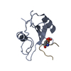
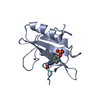
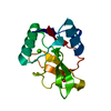
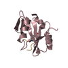
- PDB-3n8m: Crystal Structure of the Grb2 SH2 Domain in Complex with An Acycl... -
+ Open data

- Basic information
| Entry | Database: PDB / ID: 3n8m | ||||||
|---|---|---|---|---|---|---|---|
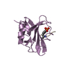
| Title | Crystal Structure of the Grb2 SH2 Domain in Complex with An Acyclic Ligand Having the Sequence pYVNVP | ||||||
 Components Components |
| ||||||
 Keywords Keywords | PROTEIN BINDING/PEPTIDE / Grb2 SH2 domain / ligand preorganization / macrocycles / macrocyclic ligands / PROTEIN BINDING-PEPTIDE complex | ||||||
| Function / homology |  Function and homology information Function and homology informationguanyl-nucleotide exchange factor adaptor activity / Grb2-EGFR complex / branching involved in labyrinthine layer morphogenesis / STAT5 Activation / Co-inhibition by BTLA / neurotrophin TRKA receptor binding / COP9 signalosome / Activated NTRK2 signals through PI3K / MET receptor recycling / transmembrane receptor protein tyrosine kinase adaptor activity ...guanyl-nucleotide exchange factor adaptor activity / Grb2-EGFR complex / branching involved in labyrinthine layer morphogenesis / STAT5 Activation / Co-inhibition by BTLA / neurotrophin TRKA receptor binding / COP9 signalosome / Activated NTRK2 signals through PI3K / MET receptor recycling / transmembrane receptor protein tyrosine kinase adaptor activity / Interleukin-15 signaling / negative regulation of natural killer cell mediated cytotoxicity / Signaling by cytosolic FGFR1 fusion mutants / MET activates PTPN11 / MET activates RAP1 and RAC1 / vesicle membrane / CD28 dependent Vav1 pathway / Signaling by LTK / Signal regulatory protein family interactions / MET activates PI3K/AKT signaling / Regulation of KIT signaling / epidermal growth factor receptor binding / natural killer cell mediated cytotoxicity / STAT5 activation downstream of FLT3 ITD mutants / PI-3K cascade:FGFR3 / PI-3K cascade:FGFR2 / PI-3K cascade:FGFR4 / PI-3K cascade:FGFR1 / GRB2:SOS provides linkage to MAPK signaling for Integrins / endodermal cell differentiation / positive regulation of actin filament polymerization / RHOU GTPase cycle / RET signaling / regulation of MAPK cascade / Interleukin-3, Interleukin-5 and GM-CSF signaling / negative regulation of epidermal growth factor receptor signaling pathway / insulin receptor substrate binding / PI3K events in ERBB2 signaling / fibroblast growth factor receptor signaling pathway / PI3K Cascade / Role of LAT2/NTAL/LAB on calcium mobilization / Interleukin receptor SHC signaling / SOS-mediated signalling / Activated NTRK3 signals through RAS / Signal attenuation / Activated NTRK2 signals through RAS / GAB1 signalosome / SHC1 events in ERBB4 signaling / RHO GTPases Activate WASPs and WAVEs / Signalling to RAS / Schwann cell development / SHC-related events triggered by IGF1R / Activated NTRK2 signals through FRS2 and FRS3 / positive regulation of Rac protein signal transduction / SHC-mediated cascade:FGFR3 / MET activates RAS signaling / ephrin receptor binding / SHC-mediated cascade:FGFR2 / SHC-mediated cascade:FGFR4 / Signaling by PDGFRA transmembrane, juxtamembrane and kinase domain mutants / Signaling by PDGFRA extracellular domain mutants / Erythropoietin activates RAS / SHC-mediated cascade:FGFR1 / Signaling by FGFR4 in disease / Signaling by CSF3 (G-CSF) / FRS-mediated FGFR3 signaling / Signaling by FLT3 ITD and TKD mutants / FRS-mediated FGFR2 signaling / FRS-mediated FGFR4 signaling / phosphotyrosine residue binding / signal transduction in response to DNA damage / FRS-mediated FGFR1 signaling / Signaling by FGFR3 in disease / Tie2 Signaling / myelination / Signaling by FGFR2 in disease / Signaling by FLT3 fusion proteins / GRB2 events in EGFR signaling / SHC1 events in EGFR signaling / FLT3 Signaling / Signaling by FGFR1 in disease / FCERI mediated Ca+2 mobilization / EGFR Transactivation by Gastrin / NCAM signaling for neurite out-growth / insulin-like growth factor receptor signaling pathway / Downstream signal transduction / GRB2 events in ERBB2 signaling / Insulin receptor signalling cascade / SHC1 events in ERBB2 signaling / Antigen activates B Cell Receptor (BCR) leading to generation of second messengers / Constitutive Signaling by Overexpressed ERBB2 / Signaling by phosphorylated juxtamembrane, extracellular and kinase domain KIT mutants / T cell activation / InlB-mediated entry of Listeria monocytogenes into host cell / B cell receptor signaling pathway / cellular response to ionizing radiation / FCGR3A-mediated phagocytosis / FCERI mediated MAPK activation / Regulation of signaling by CBL / Negative regulation of FGFR3 signaling Similarity search - Function | ||||||
| Biological species |  Homo sapiens (human) Homo sapiens (human) | ||||||
| Method |  X-RAY DIFFRACTION / MOLECULAR REPLACEMENT / Resolution: 2 Å X-RAY DIFFRACTION / MOLECULAR REPLACEMENT / Resolution: 2 Å | ||||||
 Authors Authors | Whiddon, B.B. / Clements, J.H. / Martin, S.F. | ||||||
 Citation Citation | Journal: ACS MED.CHEM.LETT. / Year: 2010 Title: Thermodynamic and Structural Effects of Macrocyclization as a Constraining Method in Protein-Ligand Interactions. Authors: Delorbe, J.E. / Clements, J.H. / Whiddon, B.B. / Martin, S.F. | ||||||
| History |
|
- Structure visualization
| Structure viewer | Molecule: MolmilJmol/JSmol |
|---|
- Downloads & links
-Download
| PDBx/mmCIF format | 3n8m.cif.gz | 40 KB | Display | PDBx/mmCIF format |
|---|---|---|---|---|
| PDB format | pdb3n8m.ent.gz | 26.2 KB | Display | PDB format |
| PDBx/mmJSON format | 3n8m.json.gz | Tree view | PDBx/mmJSON format | |
| Others |  Other downloads Other downloads |
-Validation report
| Arichive directory | https://data.pdbj.org/pub/pdb/validation_reports/n8/3n8mftp://data.pdbj.org/pub/pdb/validation_reports/n8/3n8m | HTTPS FTP |
|---|
-Related structure data
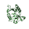
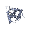
| Related structure data | 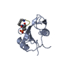 3n7yC 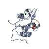 3n84C  2huwS S: Starting model for refinement C: citing same article ( |
|---|---|
| Similar structure data |


-Links
 PDBj
PDBj


- Assembly
| Deposited unit | 
| ||||||||
|---|---|---|---|---|---|---|---|---|---|
| 1 |
| ||||||||
| 2 | 
| ||||||||
| Unit cell |
|
-Components
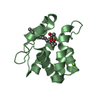
| #1: Protein | Mass: 13758.543 Da / Num. of mol.: 1 / Fragment: Grb2 SH2 domain, residues 55-153 Source method: isolated from a genetically manipulated source Details: Protein was expressed with a C-terminal six-HIS tag Source: (gene. exp.) Homo sapiens (human) / Gene: GRB2, ASH / Plasmid: pQE-60 / Production host:  |
|---|---|
| #2: Protein/peptide | Mass: 781.832 Da / Num. of mol.: 1 / Source method: obtained synthetically |
| #3: Chemical | ChemComp-GOL /   Mass: 92.094 Da / Num. of mol.: 1 / Source method: obtained synthetically / Formula: C3H8O3 Mass: 92.094 Da / Num. of mol.: 1 / Source method: obtained synthetically / Formula: C3H8O3 |
| #4: Water | ChemComp-HOH /  Mass: 18.015 Da / Num. of mol.: 107 / Source method: isolated from a natural source / Formula: H2O Mass: 18.015 Da / Num. of mol.: 107 / Source method: isolated from a natural source / Formula: H2O |
-Experimental details
-Experiment
| Experiment | Method: X-RAY DIFFRACTION / Number of used crystals: 1 |
|---|
- Sample preparation
| Crystal | Density Matthews: 1.795 Å3/Da / Density % sol: 31.48 % |
|---|---|
| Crystal grow | Temperature: 298 K / Method: vapor diffusion, hanging drop / pH: 8.5 Details: Ligand in lyophilized powder form was dissolved in a 7.2 mg/mL solution of Grb2 SH2 in water such to give a protein/ligand molar ratio of 1:1.7. 4 uL of this solution was mixed with 3 uL of ...Details: Ligand in lyophilized powder form was dissolved in a 7.2 mg/mL solution of Grb2 SH2 in water such to give a protein/ligand molar ratio of 1:1.7. 4 uL of this solution was mixed with 3 uL of 12% v/v glycerol, 1.5 M ammonium sulfate, 0.1 M TRIS, pH 8.5 to create the hanging drop, which yielded crystals of the protein-ligand complex after 8 weeks, VAPOR DIFFUSION, HANGING DROP, temperature 298K |
-Data collection
| Diffraction | Mean temperature: 100 K |
|---|---|
| Diffraction source | Source: ROTATING ANODE / Type: RIGAKU RU200 / Wavelength: 1.5418 Å |
| Detector | Type: MAR scanner 345 mm plate / Detector: IMAGE PLATE / Date: Jul 27, 2007 |
| Radiation | Monochromator: Blue max-flux confocal / Protocol: SINGLE WAVELENGTH / Monochromatic (M) / Laue (L): M / Scattering type: x-ray |
| Radiation wavelength | Wavelength: 1.5418 Å / Relative weight: 1 |
| Reflection | Resolution: 1.86→34.82 Å / Num. all: 9489 / Num. obs: 9451 / % possible obs: 99.6 % / Observed criterion σ(F): 0 / Observed criterion σ(I): 0 / Redundancy: 22.1 % / Rmerge(I) obs: 0.066 / Net I/σ(I): 60.6 |
| Reflection shell | Resolution: 1.86→1.93 Å / Redundancy: 19.5 % / Rmerge(I) obs: 0.152 / Mean I/σ(I) obs: 21.1 / Num. unique all: 912 / % possible all: 97 |
- Processing
| Software |
| |||||||||||||||||||||||||
|---|---|---|---|---|---|---|---|---|---|---|---|---|---|---|---|---|---|---|---|---|---|---|---|---|---|---|
| Refinement | Method to determine structure: MOLECULAR REPLACEMENT Starting model: 2HUW Resolution: 2→34.82 Å / Cross valid method: THROUGHOUT / σ(F): 0 / σ(I): 0 / Stereochemistry target values: Engh & Huber
| |||||||||||||||||||||||||
| Displacement parameters |
| |||||||||||||||||||||||||
| Refinement step | Cycle: LAST / Resolution: 2→34.82 Å
|
