 Movie
Movie Controller

[English] 日本語
 Yorodumi
- PDB-3n76: Crystal structure of 3-dehydroquinate dehydratase from Mycobacter... -
+ Open data

- Basic information
| Entry | Database: PDB / ID: 3n76 | ||||||
|---|---|---|---|---|---|---|---|
| Title | Crystal structure of 3-dehydroquinate dehydratase from Mycobacterium tuberculosis in complex with compound 5 | ||||||
 Components Components | 3-dehydroquinate dehydratase | ||||||
 Keywords Keywords | LYASE/LYASE INHIBITOR / dehydroquinate dehydratase / aroD / shikimate pathway / drug discovery / mycobacterium tuberculosis / LYASE / LYASE-LYASE INHIBITOR complex | ||||||
| Function / homology |  Function and homology information Function and homology informationquinate catabolic process / Chorismate via Shikimate Pathway / 3-dehydroquinate dehydratase / 3-dehydroquinate dehydratase activity / chorismate biosynthetic process / aromatic amino acid biosynthetic process / amino acid biosynthetic process / cytosol Similarity search - Function | ||||||
| Biological species |   Mycobacterium tuberculosis (bacteria) Mycobacterium tuberculosis (bacteria) | ||||||
| Method |  X-RAY DIFFRACTION / SYNCHROTRON / MOLECULAR REPLACEMENT / Resolution: 1.9 Å X-RAY DIFFRACTION / SYNCHROTRON / MOLECULAR REPLACEMENT / Resolution: 1.9 Å | ||||||
 Authors Authors | Dias, M.V.B. / Snee, W.C. / Bromfield, K.M. / Payne, R. / Palaninathan, S.K. / Ciulli, A. / Howard, N.I. / Abell, C. / Sacchettini, J.C. / Blundell, T.L. | ||||||
 Citation Citation | Journal: Biochem.J. / Year: 2011 Title: Structural investigation of inhibitor designs targeting 3-dehydroquinate dehydratase from the shikimate pathway of Mycobacterium tuberculosis. Authors: Dias, M.V. / Snee, W.C. / Bromfield, K.M. / Payne, R.J. / Palaninathan, S.K. / Ciulli, A. / Howard, N.I. / Abell, C. / Sacchettini, J.C. / Blundell, T.L. | ||||||
| History |
|
- Structure visualization
| Structure viewer | Molecule: MolmilJmol/JSmol |
|---|
- Downloads & links
-Download
| PDBx/mmCIF format | 3n76.cif.gz | 46.2 KB | Display | PDBx/mmCIF format |
|---|---|---|---|---|
| PDB format | pdb3n76.ent.gz | 32.3 KB | Display | PDB format |
| PDBx/mmJSON format | 3n76.json.gz | Tree view | PDBx/mmJSON format | |
| Others |  Other downloads Other downloads |
-Validation report
| Arichive directory | https://data.pdbj.org/pub/pdb/validation_reports/n7/3n76ftp://data.pdbj.org/pub/pdb/validation_reports/n7/3n76 | HTTPS FTP |
|---|
-Related structure data
| Related structure data |  3n59C  3n7aC  3n86C  3n87C 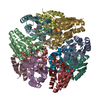 3n8kC  3n8nC  1h0rS C: citing same article ( S: Starting model for refinement |
|---|---|
| Similar structure data |


-Links
 PDBj
PDBj
- Assembly
| Deposited unit | 
| ||||||||||||
|---|---|---|---|---|---|---|---|---|---|---|---|---|---|
| 1 | x 12
| ||||||||||||
| Unit cell |
| ||||||||||||
| Components on special symmetry positions |
|
-Components
| #1: Protein | Mass: 15807.932 Da / Num. of mol.: 1 Source method: isolated from a genetically manipulated source Source: (gene. exp.) Mycobacterium tuberculosis (bacteria) / Gene: aroD, aroQ, MT2612, MTCY159.19, Rv2537c / Plasmid: pET28(a) / Production host: References: UniProt: P0A4Z6, UniProt: P9WPX7*PLUS, 3-dehydroquinate dehydratase |
|---|---|
| #2: Chemical | ChemComp-CA2 / (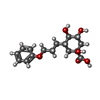  Mass: 310.342 Da / Num. of mol.: 1 / Source method: obtained synthetically / Formula: C16H22O6 Mass: 310.342 Da / Num. of mol.: 1 / Source method: obtained synthetically / Formula: C16H22O6 |
| #3: Water | ChemComp-HOH /  Mass: 18.015 Da / Num. of mol.: 161 / Source method: isolated from a natural source / Formula: H2O Mass: 18.015 Da / Num. of mol.: 161 / Source method: isolated from a natural source / Formula: H2O |
-Experimental details
-Experiment
| Experiment | Method: X-RAY DIFFRACTION / Number of used crystals: 1 |
|---|
- Sample preparation
| Crystal | Density Matthews: 2.66 Å3/Da / Density % sol: 53.8 % |
|---|---|
| Crystal grow | Temperature: 298 K / Method: vapor diffusion, hanging drop / pH: 7.5 Details: Hepes, pH7.5 peg 6000, VAPOR DIFFUSION, HANGING DROP, temperature 298K |
-Data collection
| Diffraction | Mean temperature: 100 K |
|---|---|
| Diffraction source | Source: SYNCHROTRON / Site: ESRF  / Beamline: BM14 / Wavelength: 0.97825 Å / Beamline: BM14 / Wavelength: 0.97825 Å |
| Detector | Type: MAR scanner 345 mm plate / Detector: IMAGE PLATE / Date: 2009 Details: Si(111) monochromator. Mirror 1: Grazing angle 2.8 mrad, vertical focusing. Mir ror 2: vertical and horizontal focusing. |
| Radiation | Monochromator: Si(111) / Protocol: SINGLE WAVELENGTH / Monochromatic (M) / Laue (L): M / Scattering type: x-ray |
| Radiation wavelength | Wavelength: 0.97825 Å / Relative weight: 1 |
| Reflection | Resolution: 1.9→31.6 Å / Num. all: 13318 / Num. obs: 13302 / % possible obs: 99.8 % / Observed criterion σ(F): 2 / Observed criterion σ(I): 2 / Redundancy: 4.8 % / Biso Wilson estimate: 17.725 Å2 / Rmerge(I) obs: 0.133 / Rsym value: 0.121 / Net I/σ(I): 5.5 |
| Reflection shell | Resolution: 1.9→2 Å / Redundancy: 4.6 % / Rmerge(I) obs: 0.805 / Mean I/σ(I) obs: 1 / Num. unique all: 1929 / Rsym value: 0.53 / % possible all: 98.5 |
- Processing
| Software |
| ||||||||||||||||||||||||||||||||||||||||||||||||||||||||||||||||||||||||||||||||||||||||||||||||||||||||||||||||||||||||||||||||||||||||||||||||||||||||||||||||||||||||||
|---|---|---|---|---|---|---|---|---|---|---|---|---|---|---|---|---|---|---|---|---|---|---|---|---|---|---|---|---|---|---|---|---|---|---|---|---|---|---|---|---|---|---|---|---|---|---|---|---|---|---|---|---|---|---|---|---|---|---|---|---|---|---|---|---|---|---|---|---|---|---|---|---|---|---|---|---|---|---|---|---|---|---|---|---|---|---|---|---|---|---|---|---|---|---|---|---|---|---|---|---|---|---|---|---|---|---|---|---|---|---|---|---|---|---|---|---|---|---|---|---|---|---|---|---|---|---|---|---|---|---|---|---|---|---|---|---|---|---|---|---|---|---|---|---|---|---|---|---|---|---|---|---|---|---|---|---|---|---|---|---|---|---|---|---|---|---|---|---|---|---|---|
| Refinement | Method to determine structure: MOLECULAR REPLACEMENT Starting model: 1H0R Resolution: 1.9→31.6 Å / Cor.coef. Fo:Fc: 0.945 / Cor.coef. Fo:Fc free: 0.92 / SU B: 2.987 / SU ML: 0.09 / Cross valid method: THROUGHOUT / σ(I): 2 / ESU R Free: 0.136 / Stereochemistry target values: MAXIMUM LIKELIHOOD / Details: HYDROGENS HAVE BEEN ADDED IN THE RIDING POSITIONS
| ||||||||||||||||||||||||||||||||||||||||||||||||||||||||||||||||||||||||||||||||||||||||||||||||||||||||||||||||||||||||||||||||||||||||||||||||||||||||||||||||||||||||||
| Solvent computation | Ion probe radii: 0.8 Å / Shrinkage radii: 0.8 Å / VDW probe radii: 1.4 Å / Solvent model: MASK | ||||||||||||||||||||||||||||||||||||||||||||||||||||||||||||||||||||||||||||||||||||||||||||||||||||||||||||||||||||||||||||||||||||||||||||||||||||||||||||||||||||||||||
| Displacement parameters | Biso mean: 19.439 Å2 | ||||||||||||||||||||||||||||||||||||||||||||||||||||||||||||||||||||||||||||||||||||||||||||||||||||||||||||||||||||||||||||||||||||||||||||||||||||||||||||||||||||||||||
| Refinement step | Cycle: LAST / Resolution: 1.9→31.6 Å
| ||||||||||||||||||||||||||||||||||||||||||||||||||||||||||||||||||||||||||||||||||||||||||||||||||||||||||||||||||||||||||||||||||||||||||||||||||||||||||||||||||||||||||
| Refine LS restraints |
| ||||||||||||||||||||||||||||||||||||||||||||||||||||||||||||||||||||||||||||||||||||||||||||||||||||||||||||||||||||||||||||||||||||||||||||||||||||||||||||||||||||||||||
| LS refinement shell | Resolution: 1.901→1.951 Å / Total num. of bins used: 20
|
