 Movie
Movie Controller


+ Open data

- Basic information
| Entry | Database: PDB / ID: 3n0k | ||||||
|---|---|---|---|---|---|---|---|
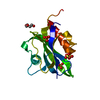
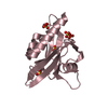
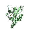
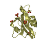
| Title | Proteinase inhibitor from Coprinopsis cinerea | ||||||
 Components Components | Serine protease inhibitor 1 | ||||||
 Keywords Keywords | Hydrolase Inhibitor / Proteinase inhibitor | ||||||
| Function / homology | Peptidase inhibitor I66 / Peptidase inhibitor I66 / Trefoil (Acidic Fibroblast Growth Factor, subunit A) - #50 / Trefoil (Acidic Fibroblast Growth Factor, subunit A) / Trefoil / serine-type endopeptidase inhibitor activity / Mainly Beta / Serine protease inhibitor 1 Function and homology information Function and homology information | ||||||
| Biological species |  Coprinopsis cinerea (fungus) Coprinopsis cinerea (fungus) | ||||||
| Method |  X-RAY DIFFRACTION / MOLECULAR REPLACEMENT / Resolution: 1.8 Å X-RAY DIFFRACTION / MOLECULAR REPLACEMENT / Resolution: 1.8 Å | ||||||
 Authors Authors | Renko, M. / Sabotic, J. / Bleuler-Martinez, S. / Kallert, S. / Avanzo, P. / Kos, J. / Aebi, M. / Kuenzler, M. / Turk, D. | ||||||
 Citation Citation | Journal: J.Biol.Chem. / Year: 2012 Title: Structural Basis of Trypsin Inhibition and Entomotoxicity of Cospin, Serine Protease Inhibitor Involved in Defense of Coprinopsis cinerea Fruiting Bodies. Authors: Sabotic, J. / Bleuler-Martinez, S. / Renko, M. / Avanzo Caglic, P. / Kallert, S. / Strukelj, B. / Turk, D. / Aebi, M. / Kos, J. / Kunzler, M. | ||||||
| History |
|
- Structure visualization
| Structure viewer | Molecule: MolmilJmol/JSmol |
|---|
- Downloads & links
-Download
| PDBx/mmCIF format | 3n0k.cif.gz | 48.1 KB | Display | PDBx/mmCIF format |
|---|---|---|---|---|
| PDB format | pdb3n0k.ent.gz | 33.9 KB | Display | PDB format |
| PDBx/mmJSON format | 3n0k.json.gz | Tree view | PDBx/mmJSON format | |
| Others |  Other downloads Other downloads |
-Validation report
| Arichive directory | https://data.pdbj.org/pub/pdb/validation_reports/n0/3n0kftp://data.pdbj.org/pub/pdb/validation_reports/n0/3n0k | HTTPS FTP |
|---|
-Related structure data
| Similar structure data |
|---|


-Links
 PDBj
PDBj- Assembly
| Deposited unit | 
| ||||||||
|---|---|---|---|---|---|---|---|---|---|
| 1 |
| ||||||||
| Unit cell |
| ||||||||
| Components on special symmetry positions |
|
-Components
| #1: Protein | Mass: 16730.748 Da / Num. of mol.: 1 Source method: isolated from a genetically manipulated source Source: (gene. exp.) Coprinopsis cinerea (fungus) / Plasmid: pet24 / Production host:  |
|---|---|
| #2: Water | ChemComp-HOH /  Mass: 18.015 Da / Num. of mol.: 251 / Source method: isolated from a natural source / Formula: H2O Mass: 18.015 Da / Num. of mol.: 251 / Source method: isolated from a natural source / Formula: H2O |
-Experimental details
-Experiment
| Experiment | Method: X-RAY DIFFRACTION / Number of used crystals: 1 |
|---|
- Sample preparation
| Crystal | Density Matthews: 2.45 Å3/Da / Density % sol: 49.87 % |
|---|---|
| Crystal grow | Temperature: 293 K / Method: vapor diffusion, sitting drop / pH: 6 Details: 0.1 M MES buffer, 20% MPD, pH 6.0, VAPOR DIFFUSION, SITTING DROP, temperature 293K |
-Data collection
| Diffraction | Mean temperature: 100 K |
|---|---|
| Diffraction source | Source: ROTATING ANODE / Type: RIGAKU RU200 / Wavelength: 1.54 Å |
| Detector | Type: MAR scanner 345 mm plate / Detector: IMAGE PLATE / Date: Oct 10, 2009 / Details: mirrors |
| Radiation | Monochromator: YALE MIRRORS / Protocol: SINGLE WAVELENGTH / Monochromatic (M) / Laue (L): M / Scattering type: x-ray |
| Radiation wavelength | Wavelength: 1.54 Å / Relative weight: 1 |
| Reflection | Resolution: 1.8→16.87 Å / Num. all: 15292 / Num. obs: 14500 / % possible obs: 95 % / Observed criterion σ(F): 1 / Observed criterion σ(I): 1 / Redundancy: 7.1 % / Rmerge(I) obs: 0.034 / Χ2: 2.396 / Net I/σ(I): 51.3 |
| Reflection shell | Resolution: 1.8→1.83 Å / Redundancy: 3.8 % / Rmerge(I) obs: 0.115 / Num. unique all: 621 / Χ2: 3.508 / % possible all: 83.4 |
- Processing
| Software |
| |||||||||||||||||||||||||||||||||||||||||||||||||||||||||||||||||
|---|---|---|---|---|---|---|---|---|---|---|---|---|---|---|---|---|---|---|---|---|---|---|---|---|---|---|---|---|---|---|---|---|---|---|---|---|---|---|---|---|---|---|---|---|---|---|---|---|---|---|---|---|---|---|---|---|---|---|---|---|---|---|---|---|---|---|
| Refinement | Method to determine structure: MOLECULAR REPLACEMENT / Resolution: 1.8→16.87 Å / Cor.coef. Fo:Fc: 0.961 / Cor.coef. Fo:Fc free: 0.952 / WRfactor Rfree: 0.215 / WRfactor Rwork: 0.185 / Occupancy max: 1 / Occupancy min: 0 / FOM work R set: 0.88 / SU B: 2.207 / SU ML: 0.071 / SU R Cruickshank DPI: 0.133 / SU Rfree: 0.116 / Cross valid method: THROUGHOUT / σ(F): 0 / ESU R Free: 0.116 / Stereochemistry target values: MAXIMUM LIKELIHOOD Details: HYDROGENS HAVE BEEN ADDED IN THE RIDING POSITIONS U VALUES: REFINED INDIVIDUALLY
| |||||||||||||||||||||||||||||||||||||||||||||||||||||||||||||||||
| Solvent computation | Ion probe radii: 0.8 Å / Shrinkage radii: 0.8 Å / VDW probe radii: 1.4 Å / Solvent model: MASK | |||||||||||||||||||||||||||||||||||||||||||||||||||||||||||||||||
| Displacement parameters | Biso max: 66.86 Å2 / Biso mean: 23.282 Å2 / Biso min: 8.41 Å2
| |||||||||||||||||||||||||||||||||||||||||||||||||||||||||||||||||
| Refinement step | Cycle: LAST / Resolution: 1.8→16.87 Å
| |||||||||||||||||||||||||||||||||||||||||||||||||||||||||||||||||
| Refine LS restraints |
| |||||||||||||||||||||||||||||||||||||||||||||||||||||||||||||||||
| LS refinement shell | Resolution: 1.8→1.845 Å / Total num. of bins used: 20
|
