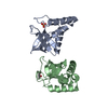
- PDB-3mw8: Crystal structure of an UROPORPHYRINOGEN-III SYNTHASE (Sama_3255)... -
+
データを開く
IDまたはキーワード:
読み込み中...
-
基本情報
登録情報
データベース: PDB / ID: 3mw8
タイトル
Crystal structure of an UROPORPHYRINOGEN-III SYNTHASE (Sama_3255) from SHEWANELLA AMAZONENSIS SB2B at 1.65 A resolution
要素
Uroporphyrinogen-III synthase
キーワード
LYASE / Structural Genomics / Joint Center for Structural Genomics / JCSG / Protein Structure Initiative / PSI-2 / HemD-like / heme
機能・相同性
機能・相同性情報
uroporphyrinogen-III synthase / uroporphyrinogen-III synthase activity / uroporphyrinogen III biosynthetic process / protoporphyrinogen IX biosynthetic process 類似検索 - 分子機能
THE CONSTRUCT WAS EXPRESSED WITH AN N-TERMINAL PURIFICATION TAG MGSDKIHHHHHHENLYFQG. CLEAVAGE OF ...THE CONSTRUCT WAS EXPRESSED WITH AN N-TERMINAL PURIFICATION TAG MGSDKIHHHHHHENLYFQG. CLEAVAGE OF THE TAG WITH TEV PROTEASE LEAVES ONLY A GLYCINE (0) FOLLOWED BY THE TARGET SEQUENCE. HOWEVER, THE CLEAVAGE OF THE TAG WAS INCOMPLETE LEAVING A MIXTURE OF PROTEIN WITH THE INTACT TAG AND CLEAVED TAG.
モノクロメーター: Double crystal monochromator / プロトコル: SINGLE WAVELENGTH / 単色(M)・ラウエ(L): M / 散乱光タイプ: x-ray
放射波長
波長: 0.97927 Å / 相対比: 1
反射
解像度: 1.65→38.525 Å / Num. obs: 35551 / % possible obs: 99.2 % / Observed criterion σ(I): -3 / Biso Wilson estimate: 25.002 Å2 / Rmerge(I) obs: 0.048 / Net I/σ(I): 13.48
反射 シェル
解像度 (Å)
Rmerge(I) obs
Mean I/σ(I) obs
Num. measured obs
Num. unique obs
% possible all
1.65-1.71
0.442
2.4
13207
3628
99.8
1.71-1.78
0.299
3.5
13266
3595
100
1.78-1.86
0.213
4.9
12945
3486
99.8
1.86-1.96
0.147
7.1
13286
3600
99.9
1.96-2.08
0.092
10.6
12881
3485
99.9
2.08-2.24
0.068
14.4
13169
3557
99.8
2.24-2.46
0.056
17.8
12856
3476
99.5
2.46-2.82
0.05
20.9
13189
3618
99.5
2.82-3.55
0.039
25.5
12925
3547
98.5
3.55-38.525
0.036
27.8
12299
3550
95.8
-
位相決定
位相決定
手法: 単波長異常分散
-
解析
ソフトウェア
名称
バージョン
分類
NB
REFMAC
5.5.0109
精密化
PHENIX
精密化
SHELX
位相決定
MolProbity
3beta29
モデル構築
XSCALE
データスケーリング
PDB_EXTRACT
3.006
データ抽出
XDS
データ削減
SHELXD
位相決定
autoSHARP
位相決定
精密化
構造決定の手法: 単波長異常分散 / 解像度: 1.65→38.525 Å / Cor.coef. Fo:Fc: 0.966 / Cor.coef. Fo:Fc free: 0.959 / Occupancy max: 1 / Occupancy min: 0.3 / SU B: 3.519 / SU ML: 0.058 / 交差検証法: THROUGHOUT / σ(F): 0 / ESU R Free: 0.086 立体化学のターゲット値: MAXIMUM LIKELIHOOD WITH PHASES 詳細: 1. HYDROGENS HAVE BEEN ADDED IN THE RIDING POSITIONS. 2. A MET-INHIBITION PROTOCOL WAS USED FOR SELENOMETHIONINE INCORPORATION DURING PROTEIN EXPRESSION. THE OCCUPANCY OF THE SE ATOMS IN THE ...詳細: 1. HYDROGENS HAVE BEEN ADDED IN THE RIDING POSITIONS. 2. A MET-INHIBITION PROTOCOL WAS USED FOR SELENOMETHIONINE INCORPORATION DURING PROTEIN EXPRESSION. THE OCCUPANCY OF THE SE ATOMS IN THE MSE RESIDUES WAS REDUCED TO 0.75 FOR THE REDUCED SCATTERING POWER DUE TO PARTIAL S-MET INCORPORATION. 3. ATOM RECORD CONTAINS SUM OF TLS AND RESIDUAL B FACTORS. ANISOU RECORD CONTAINS SUM OF TLS AND RESIDUAL U FACTORS. 4. ETHYLENE GLYCOL (EDO) MODELED ARE PRESENT CRYO SOLUTION.
Rfactor
反射数
%反射
Selection details
Rfree
0.203
1814
5.1 %
RANDOM
Rwork
0.175
-
-
-
obs
0.177
35537
99.26 %
-
溶媒の処理
イオンプローブ半径: 0.8 Å / 減衰半径: 0.8 Å / VDWプローブ半径: 1.2 Å / 溶媒モデル: MASK
 ムービー
ムービー コントローラー


 データを開く
 基本情報
基本情報 要素
要素 キーワード
キーワード 機能・相同性情報
 Shewanella amazonensis (バクテリア)
 X線回折 /
  データ登録者
 引用
引用 構造の表示
構造の表示 ダウンロードとリンク
 その他のダウンロード


 PDBj
PDBj 集合体
集合体

 分子量: 62.068 Da / 分子数: 12 / 由来タイプ: 合成 / 式: C2H6O2
 分子量: 18.015 Da / 分子数: 226 / 由来タイプ: 天然 / 式: H2O
 試料調製
試料調製 / ビームライン: BL9-2 / 波長: 0.97927
  解析
解析