- PDB-3mp7: Lateral opening of a translocon upon entry of protein suggests th... -
+
Open data
ID or keywords:
Loading...
-
Basic information
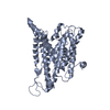
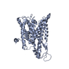
Entry
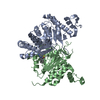
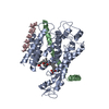
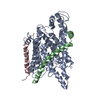
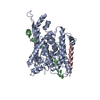
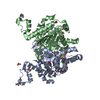
Database: PDB / ID: 3mp7
Title
Lateral opening of a translocon upon entry of protein suggests the mechanism of insertion into membranes
Components
Preprotein translocase subunit secE
Preprotein translocase subunit secY
Keywords
PROTEIN TRANSPORT / Membrane Protein Complex / Preprotein Translocase / Membrane Insertion / Structural Genomics / PSI-2 / Protein Structure Initiative / Center for Structures of Membrane Proteins / CSMP
Function / homology
Function and homology information
intracellular protein transmembrane transport / protein secretion / transmembrane protein transporter activity / protein targeting / plasma membrane Similarity search - Function
Preprotein translocase SecE subunit / Preprotein translocase SecY subunit / SecY subunit domain / Protein translocase subunit SecY / Protein translocase SEC61 complex, gamma subunit / Protein translocase SecE domain superfamily / Translocon Sec61/SecY, plug domain / Plug domain of Sec61p / Protein secY signature 1. / Protein secY signature 2. ...Preprotein translocase SecE subunit / Preprotein translocase SecY subunit / SecY subunit domain / Protein translocase subunit SecY / Protein translocase SEC61 complex, gamma subunit / Protein translocase SecE domain superfamily / Translocon Sec61/SecY, plug domain / Plug domain of Sec61p / Protein secY signature 1. / Protein secY signature 2. / Protein translocase complex, SecE/Sec61-gamma subunit / SecY/SEC61-alpha family / SecY domain superfamily / SecY conserved site / SecY / Single alpha-helices involved in coiled-coils or other helix-helix interfaces / Up-down Bundle / Orthogonal Bundle / Mainly Alpha Similarity search - Domain/homology
PreproteintranslocasesubunitsecY / Protein transport protein SEC61 subunit alpha homolog
Mass: 53939.324 Da / Num. of mol.: 1 Source method: isolated from a genetically manipulated source Source: (gene. exp.) Pyrococcus furiosus (archaea) / Gene: secY / Plasmid: pBAD / Production host: Escherichia coli (E. coli) / Strain (production host): BL21(AI) / References: UniProt: Q8U019
#2: Protein
PreproteintranslocasesubunitsecE / Protein transport protein Sec61 gamma subunit homolog
Mass: 6944.483 Da / Num. of mol.: 1 Source method: isolated from a genetically manipulated source Source: (gene. exp.) Pyrococcus furiosus (archaea) / Gene: secE / Plasmid: pBAD / Production host: Escherichia coli (E. coli) / Strain (production host): BL21(AI) / References: UniProt: Q8TZK2
-
Experimental details
-
Experiment
Experiment
Method: X-RAY DIFFRACTION / Number of used crystals: 2
-
Sample preparation
Crystal
Density Matthews: 5 Å3/Da / Density % sol: 75.4 %
Crystal grow
Temperature: 273 K / Method: vapor diffusion, hanging drop / pH: 6.2 Details: PEG4000-PEG8000 15-25%, MES 100mM pH 6.2, Ca or Mg Acetate 50-200mM, VAPOR DIFFUSION, HANGING DROP, temperature 273K
In the structure databanks used in Yorodumi, some data are registered as the other names, "COVID-19 virus" and "2019-nCoV". Here are the details of the virus and the list of structure data.
Jan 31, 2019. EMDB accession codes are about to change! (news from PDBe EMDB page)
EMDB accession codes are about to change! (news from PDBe EMDB page)
The allocation of 4 digits for EMDB accession codes will soon come to an end. Whilst these codes will remain in use, new EMDB accession codes will include an additional digit and will expand incrementally as the available range of codes is exhausted. The current 4-digit format prefixed with “EMD-” (i.e. EMD-XXXX) will advance to a 5-digit format (i.e. EMD-XXXXX), and so on. It is currently estimated that the 4-digit codes will be depleted around Spring 2019, at which point the 5-digit format will come into force.
The EM Navigator/Yorodumi systems omit the EMD- prefix.
Related info.:Q: What is EMD? / ID/Accession-code notation in Yorodumi/EM Navigator
Yorodumi is a browser for structure data from EMDB, PDB, SASBDB, etc.
This page is also the successor to EM Navigator detail page, and also detail information page/front-end page for Omokage search.
The word "yorodu" (or yorozu) is an old Japanese word meaning "ten thousand". "mi" (miru) is to see.
Related info.:EMDB / PDB / SASBDB / Comparison of 3 databanks / Yorodumi Search / Aug 31, 2016. New EM Navigator & Yorodumi / Yorodumi Papers / Jmol/JSmol / Function and homology information / Changes in new EM Navigator and Yorodumi
 Movie
Movie Controller

 Yorodumi
 Open data
 Basic information
 Components
 Keywords
 Function and homology information
 Pyrococcus furiosus (archaea)
 X-RAY DIFFRACTION /
  Authors
 Citation
 Structure visualization
 Downloads & links
 Other downloads


 PDBj
PDBj
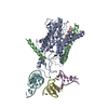
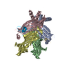
 Assembly
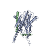

 Sample preparation
 Processing