 Movie
Movie Controller

[English] 日本語
 Yorodumi
- PDB-3mn9: Structures of actin-bound WH2 domains of Spire and the implicatio... -
+ Open data

- Basic information
| Entry | Database: PDB / ID: 3mn9 | ||||||
|---|---|---|---|---|---|---|---|
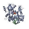
| Title | Structures of actin-bound WH2 domains of Spire and the implication for filament nucleation | ||||||
 Components Components |
| ||||||
 Keywords Keywords | Contractile Protein/Protein binding / WH2 domain / Spire / Actin complex / Contractile Protein-Protein binding complex | ||||||
| Function / homology |  Function and homology information Function and homology informationchorion-containing eggshell formation / pole plasm RNA localization / Gap junction degradation / Formation of annular gap junctions / EPHB-mediated forward signaling / EPH-ephrin mediated repulsion of cells / Cell-extracellular matrix interactions / RHOBTB2 GTPase cycle / RHOF GTPase cycle / VEGFA-VEGFR2 Pathway ...chorion-containing eggshell formation / pole plasm RNA localization / Gap junction degradation / Formation of annular gap junctions / EPHB-mediated forward signaling / EPH-ephrin mediated repulsion of cells / Cell-extracellular matrix interactions / RHOBTB2 GTPase cycle / RHOF GTPase cycle / VEGFA-VEGFR2 Pathway / ovarian fusome organization / oocyte karyosome formation / Platelet degranulation / MAP2K and MAPK activation / establishment of meiotic spindle localization / RHO GTPases Activate WASPs and WAVEs / Regulation of actin dynamics for phagocytic cup formation / actin filament-based process / pole plasm oskar mRNA localization / DNA Damage Recognition in GG-NER / pole plasm assembly / Clathrin-mediated endocytosis / polar body extrusion after meiotic divisions / actin filament network formation / sperm individualization / UCH proteinases / actin nucleation / Golgi vesicle transport / maintenance of protein location in cell / cleavage furrow formation / brahma complex / tube formation / Ino80 complex / oogenesis / positive regulation of mitochondrial fission / regulation of cytoskeleton organization / mitotic cytokinesis / intracellular transport / vesicle-mediated transport / cytoplasmic vesicle membrane / actin filament organization / Hydrolases; Acting on acid anhydrides; Acting on acid anhydrides to facilitate cellular and subcellular movement / protein transport / actin cytoskeleton / actin binding / actin cytoskeleton organization / cell cortex / microtubule binding / mitochondrial outer membrane / cytoskeleton / hydrolase activity / chromatin remodeling / perinuclear region of cytoplasm / ATP binding / plasma membrane / cytoplasm Similarity search - Function | ||||||
| Biological species |  | ||||||
| Method |  X-RAY DIFFRACTION / SYNCHROTRON / MOLECULAR REPLACEMENT / Resolution: 2 Å X-RAY DIFFRACTION / SYNCHROTRON / MOLECULAR REPLACEMENT / Resolution: 2 Å | ||||||
 Authors Authors | Ducka, A.M. / Sitar, T. / Popowicz, G.M. / Huber, R. / Holak, T.A. | ||||||
 Citation Citation | Journal: Proc.Natl.Acad.Sci.USA / Year: 2010 Title: Structures of actin-bound Wiskott-Aldrich syndrome protein homology 2 (WH2) domains of Spire and the implication for filament nucleation. Authors: Ducka, A.M. / Joel, P. / Popowicz, G.M. / Trybus, K.M. / Schleicher, M. / Noegel, A.A. / Huber, R. / Holak, T.A. / Sitar, T. | ||||||
| History |
|
- Structure visualization
| Structure viewer | Molecule: MolmilJmol/JSmol |
|---|
- Downloads & links
-Download
| PDBx/mmCIF format | 3mn9.cif.gz | 92 KB | Display | PDBx/mmCIF format |
|---|---|---|---|---|
| PDB format | pdb3mn9.ent.gz | 68.1 KB | Display | PDB format |
| PDBx/mmJSON format | 3mn9.json.gz | Tree view | PDBx/mmJSON format | |
| Others |  Other downloads Other downloads |
-Validation report
| Summary document | 3mn9_validation.pdf.gz | 798.1 KB | Display | wwPDB validaton report |
|---|---|---|---|---|
| Full document | 3mn9_full_validation.pdf.gz | 801 KB | Display | |
| Data in XML | 3mn9_validation.xml.gz | 16.8 KB | Display | |
| Data in CIF | 3mn9_validation.cif.gz | 23.9 KB | Display | |
| Arichive directory | https://data.pdbj.org/pub/pdb/validation_reports/mn/3mn9ftp://data.pdbj.org/pub/pdb/validation_reports/mn/3mn9 | HTTPS FTP |
-Related structure data
| Related structure data | 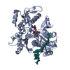 3mmvC  3mn5C 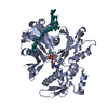 3mn6C  3mn7C 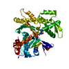 2hf4S S: Starting model for refinement C: citing same article ( |
|---|---|
| Similar structure data |


-Links
 PDBj
PDBj


- Assembly
| Deposited unit | 
| ||||||||
|---|---|---|---|---|---|---|---|---|---|
| 1 |
| ||||||||
| Unit cell |
|
-Components
| #1: Protein | Mass: 41723.527 Da / Num. of mol.: 1 / Mutation: A204E, P243K Source method: isolated from a genetically manipulated source Source: (gene. exp.)  Spodoptera frugiperda (fall armyworm) / References: UniProt: P10987 Spodoptera frugiperda (fall armyworm) / References: UniProt: P10987 |
|---|---|
| #2: Protein/peptide | Mass: 1635.006 Da / Num. of mol.: 1 / Fragment: UNP residues 371-485 Source method: isolated from a genetically manipulated source Source: (gene. exp.)  |
| #3: Chemical | ChemComp-ATP /   Mass: 507.181 Da / Num. of mol.: 1 / Source method: obtained synthetically / Formula: C10H16N5O13P3 / Comment: ATP, energy-carrying molecule*YM Mass: 507.181 Da / Num. of mol.: 1 / Source method: obtained synthetically / Formula: C10H16N5O13P3 / Comment: ATP, energy-carrying molecule*YM |
| #4: Chemical | ChemComp-CA /   Mass: 40.078 Da / Num. of mol.: 1 / Source method: obtained synthetically / Formula: Ca Mass: 40.078 Da / Num. of mol.: 1 / Source method: obtained synthetically / Formula: Ca |
| #5: Water | ChemComp-HOH /  Mass: 18.015 Da / Num. of mol.: 138 / Source method: isolated from a natural source / Formula: H2O Mass: 18.015 Da / Num. of mol.: 138 / Source method: isolated from a natural source / Formula: H2O |
| Sequence details | THE SEQUENCE OF CHAIN X IS : VQVIDELRRG VRLKKSNHER TPIEYELTPY EILMGDIRAK KYQLRKVMVN GDIPPRVKKD ...THE SEQUENCE OF CHAIN X IS : VQVIDELRRG |
-Experimental details
-Experiment
| Experiment | Method: X-RAY DIFFRACTION / Number of used crystals: 1 |
|---|
- Sample preparation
| Crystal | Density Matthews: 2.98 Å3/Da / Density % sol: 58.77 % |
|---|---|
| Crystal grow | Temperature: 277 K / Method: vapor diffusion, sitting drop / pH: 6.6 Details: 0.2M Ammonium formate pH 6.6 20% PEG 3350, VAPOR DIFFUSION, SITTING DROP, temperature 277K |
-Data collection
| Diffraction | Mean temperature: 90 K |
|---|---|
| Diffraction source | Source: SYNCHROTRON / Site: SLS  / Beamline: X10SA / Wavelength: 0.9792 Å / Beamline: X10SA / Wavelength: 0.9792 Å |
| Detector | Type: MAR CCD 165 mm / Detector: CCD / Date: Mar 27, 2010 / Details: LN2 cooled fixed-exit Si(111) monochromator |
| Radiation | Monochromator: mirror / Protocol: SINGLE WAVELENGTH / Monochromatic (M) / Laue (L): M / Scattering type: x-ray |
| Radiation wavelength | Wavelength: 0.9792 Å / Relative weight: 1 |
| Reflection | Resolution: 2→50 Å / Num. all: 29535 / Num. obs: 22506 / % possible obs: 76.2 % / Observed criterion σ(F): 2 / Observed criterion σ(I): 2 / Rmerge(I) obs: 0.067 |
| Reflection shell | Resolution: 2→2.3 Å / Rmerge(I) obs: 0.269 / Mean I/σ(I) obs: 3.9 / % possible all: 38.5 |
- Processing
| Software |
| |||||||||||||||||||||||||||||||||||||||||||||||||||||||||||||||||
|---|---|---|---|---|---|---|---|---|---|---|---|---|---|---|---|---|---|---|---|---|---|---|---|---|---|---|---|---|---|---|---|---|---|---|---|---|---|---|---|---|---|---|---|---|---|---|---|---|---|---|---|---|---|---|---|---|---|---|---|---|---|---|---|---|---|---|
| Refinement | Method to determine structure: MOLECULAR REPLACEMENT Starting model: 2HF4 Resolution: 2→50 Å / Cor.coef. Fo:Fc: 0.954 / Cor.coef. Fo:Fc free: 0.905 / SU B: 4.951 / SU ML: 0.137 / Cross valid method: THROUGHOUT / ESU R: 0.262 / ESU R Free: 0.229 / Stereochemistry target values: MAXIMUM LIKELIHOOD / Details: HYDROGENS HAVE BEEN ADDED IN THE RIDING POSITIONS
| |||||||||||||||||||||||||||||||||||||||||||||||||||||||||||||||||
| Solvent computation | Ion probe radii: 0.8 Å / Shrinkage radii: 0.8 Å / VDW probe radii: 1.4 Å / Solvent model: MASK | |||||||||||||||||||||||||||||||||||||||||||||||||||||||||||||||||
| Displacement parameters | Biso mean: 42.81 Å2
| |||||||||||||||||||||||||||||||||||||||||||||||||||||||||||||||||
| Refinement step | Cycle: LAST / Resolution: 2→50 Å
| |||||||||||||||||||||||||||||||||||||||||||||||||||||||||||||||||
| Refine LS restraints |
| |||||||||||||||||||||||||||||||||||||||||||||||||||||||||||||||||
| LS refinement shell | Resolution: 2→2.052 Å / Total num. of bins used: 20
|
