Resolution: 2.1→19.75 Å / Cor.coef. Fo:Fc: 0.945 / Cor.coef. Fo:Fc free: 0.938 / SU B: 10.716 / SU ML: 0.146 / Cross valid method: THROUGHOUT / ESU R: 0.263 / ESU R Free: 0.19 / Stereochemistry target values: MAXIMUM LIKELIHOOD / Details: HYDROGENS HAVE BEEN ADDED IN THE RIDING POSITIONS
Rfactor
Num. reflection
% reflection
Selection details
Rfree
0.23302
2286
5 %
RANDOM
Rwork
0.20786
-
-
-
obs
0.20913
43424
100 %
-
all
-
45824
-
-
Solvent computation
Ion probe radii: 0.8 Å / Shrinkage radii: 0.8 Å / VDW probe radii: 1.4 Å / Solvent model: MASK
Displacement parameters
Biso mean: 22.757 Å2
Baniso -1
Baniso -2
Baniso -3
1-
-0.42 Å2
0 Å2
-0.71 Å2
2-
-
0.79 Å2
0 Å2
3-
-
-
-0.68 Å2
Refinement step
Cycle: LAST / Resolution: 2.1→19.75 Å
Protein
Nucleic acid
Ligand
Solvent
Total
Num. atoms
5968
0
146
204
6318
Refine LS restraints
Refine-ID
Type
Dev ideal
Dev ideal target
Number
X-RAY DIFFRACTION
r_bond_refined_d
0.006
0.021
6261
X-RAY DIFFRACTION
r_angle_refined_deg
0.884
2.013
8552
X-RAY DIFFRACTION
r_dihedral_angle_1_deg
4.162
5
781
X-RAY DIFFRACTION
r_dihedral_angle_2_deg
32.665
22.724
279
X-RAY DIFFRACTION
r_dihedral_angle_3_deg
13.376
15
966
X-RAY DIFFRACTION
r_dihedral_angle_4_deg
10.39
15
68
X-RAY DIFFRACTION
r_chiral_restr
0.055
0.2
962
X-RAY DIFFRACTION
r_gen_planes_refined
0.003
0.021
4801
X-RAY DIFFRACTION
r_mcbond_it
0.152
1.5
3888
X-RAY DIFFRACTION
r_mcangle_it
0.287
2
6281
X-RAY DIFFRACTION
r_scbond_it
0.408
3
2373
X-RAY DIFFRACTION
r_scangle_it
0.691
4.5
2269
LS refinement shell
Resolution: 2.1→2.154 Å / Total num. of bins used: 20
Rfactor
Num. reflection
% reflection
Rfree
0.293
164
-
Rwork
0.242
3123
-
obs
-
3123
100 %
Refinement TLS params.
Method: refined / Refine-ID: X-RAY DIFFRACTION
ID
L11 (°2)
L12 (°2)
L13 (°2)
L22 (°2)
L23 (°2)
L33 (°2)
S11 (Å °)
S12 (Å °)
S13 (Å °)
S21 (Å °)
S22 (Å °)
S23 (Å °)
S31 (Å °)
S32 (Å °)
S33 (Å °)
T11 (Å2)
T12 (Å2)
T13 (Å2)
T22 (Å2)
T23 (Å2)
T33 (Å2)
Origin x (Å)
Origin y (Å)
Origin z (Å)
1
2.8278
-0.3323
0.7995
1.8684
-0.9827
3.3343
-0.1301
-0.2352
0.3578
0.0614
0.0885
0.144
-0.0306
-0.3323
0.0416
0.0186
-0.011
-0.0042
0.1708
-0.0387
0.0693
37.428
12.8583
41.2618
2
6.098
-0.956
0.7572
7.0938
-1.669
5.7556
-0.079
-0.5633
-0.6866
-0.211
-0.0654
0.2283
0.9498
-0.3608
0.1444
0.2828
-0.0993
0.0385
0.2278
0.0355
0.0943
42.5656
-5.7861
45.3629
3
8.3709
2.5778
1.417
11.0585
2.4529
4.0958
-0.157
-0.7927
0.1293
1.1024
0.1236
-0.6514
0.1735
0.2092
0.0334
0.1899
0.0702
-0.0787
0.3009
0.0065
0.0601
51.5162
1.9773
60.1704
4
2.1208
-0.3131
-0.0911
2.1637
-1.0739
3.5184
-0.1291
0.2038
0.3413
-0.2011
0.1338
0.0728
0.0478
-0.2144
-0.0046
0.0344
-0.0442
-0.0307
0.2007
-0.0038
0.0753
41.842
12.7257
33.3622
5
9.1821
-2.4556
-0.8241
4.1857
0.8048
3.5913
0.0531
0.7308
-1.0873
-0.8978
-0.0721
0.389
0.6844
-0.2021
0.019
0.5119
-0.161
-0.0695
0.2518
-0.0267
0.1529
39.5025
-3.8159
26.8996
6
2.4497
1.7118
0.1893
1.8714
0.4437
3.9173
-0.2715
0.1932
-0.0805
-0.1278
0.0706
-0.4435
-0.0616
0.7599
0.2009
0.117
-0.0629
0.0004
0.2311
0.0743
0.2935
29.9457
13.2467
11.3165
7
2.8204
1.026
1.3147
2.2283
0.8412
2.4435
-0.1728
0.3667
0.1697
-0.1054
0.0242
0.1416
-0.0923
0.188
0.1486
0.0804
-0.0105
0.0369
0.0562
0.026
0.1103
11.1417
6.0498
3.1881
8
4.2294
0.2171
0.7562
2.8683
0.4076
3.333
0.1685
0.3283
-0.4027
-0.1937
-0.0382
0.0731
0.2966
0.2859
-0.1303
0.1258
0.0436
-0.0052
0.0479
-0.0547
0.1397
9.165
-9.2998
-1.4069
9
2.0967
0.3681
1.0744
1.9059
0.701
2.6426
-0.099
-0.0959
0.307
0.1396
-0.0767
0.0987
-0.1489
-0.0031
0.1756
0.0708
-0.0161
0.0129
0.0167
-0.0034
0.1192
12.5773
10.448
13.7617
10
7.2033
1.871
-0.2066
3.6568
-0.9373
4.422
0.4175
-0.7724
-0.5171
0.7764
-0.3198
-0.1641
0.486
0.1813
-0.0977
0.4157
-0.0786
-0.0145
0.0987
0.0353
0.1105
14.5222
-4.5717
23.0907
Refinement TLS group
ID
Refine-ID
Refine TLS-ID
Auth asym-ID
Auth seq-ID
1
X-RAY DIFFRACTION
1
A
6 - 135
2
X-RAY DIFFRACTION
2
A
136 - 190
3
X-RAY DIFFRACTION
3
A
191 - 224
4
X-RAY DIFFRACTION
4
A
225 - 358
5
X-RAY DIFFRACTION
5
A
359 - 396
6
X-RAY DIFFRACTION
6
B
6 - 44
7
X-RAY DIFFRACTION
7
B
45 - 138
8
X-RAY DIFFRACTION
8
B
139 - 227
9
X-RAY DIFFRACTION
9
B
228 - 357
10
X-RAY DIFFRACTION
10
B
358 - 396
+
About Yorodumi
-
News
-
Feb 9, 2022. New format data for meta-information of EMDB entries
New format data for meta-information of EMDB entries
Version 3 of the EMDB header file is now the official format.
The previous official version 1.9 will be removed from the archive.
In the structure databanks used in Yorodumi, some data are registered as the other names, "COVID-19 virus" and "2019-nCoV". Here are the details of the virus and the list of structure data.
Jan 31, 2019. EMDB accession codes are about to change! (news from PDBe EMDB page)
EMDB accession codes are about to change! (news from PDBe EMDB page)
The allocation of 4 digits for EMDB accession codes will soon come to an end. Whilst these codes will remain in use, new EMDB accession codes will include an additional digit and will expand incrementally as the available range of codes is exhausted. The current 4-digit format prefixed with “EMD-” (i.e. EMD-XXXX) will advance to a 5-digit format (i.e. EMD-XXXXX), and so on. It is currently estimated that the 4-digit codes will be depleted around Spring 2019, at which point the 5-digit format will come into force.
The EM Navigator/Yorodumi systems omit the EMD- prefix.
Related info.:Q: What is EMD? / ID/Accession-code notation in Yorodumi/EM Navigator
Yorodumi is a browser for structure data from EMDB, PDB, SASBDB, etc.
This page is also the successor to EM Navigator detail page, and also detail information page/front-end page for Omokage search.
The word "yorodu" (or yorozu) is an old Japanese word meaning "ten thousand". "mi" (miru) is to see.
Related info.:EMDB / PDB / SASBDB / Comparison of 3 databanks / Yorodumi Search / Aug 31, 2016. New EM Navigator & Yorodumi / Yorodumi Papers / Jmol/JSmol / Function and homology information / Changes in new EM Navigator and Yorodumi
 Movie
Movie Controller

 Yorodumi
 Open data
 Basic information
 Components
 Keywords
 Function and homology information
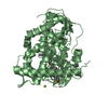
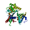
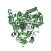
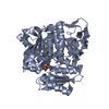
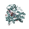
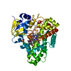
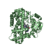
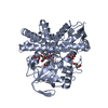
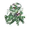
 Amycolatopsis balhimycina (bacteria)
 X-RAY DIFFRACTION /
  Authors
 Citation
 Structure visualization
 Downloads & links
 Other downloads
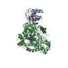


 PDBj
PDBj
 Assembly

 Mass: 616.487 Da / Num. of mol.: 2 / Source method: obtained synthetically / Formula: C34H32FeN4O4
 Mass: 92.094 Da / Num. of mol.: 10 / Source method: obtained synthetically / Formula: C3H8O3
 Mass: 18.015 Da / Num. of mol.: 204 / Source method: isolated from a natural source / Formula: H2O
 Sample preparation
 / Beamline: X10SA / Wavelength: 0.99986 Å
 Processing