 Movie
Movie Controller

[English] 日本語
 Yorodumi
- PDB-3mfb: Crystal Structure of the S-type Pyocin domain of ECA1669 protein ... -
+ Open data

- Basic information
| Entry | Database: PDB / ID: 3mfb | ||||||
|---|---|---|---|---|---|---|---|
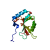
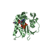
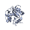
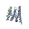
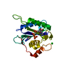
| Title | Crystal Structure of the S-type Pyocin domain of ECA1669 protein from Erwinia carotovora, Northeast Structural Genomics Consortium Target EwR82C | ||||||
 Components Components | Uncharacterized protein | ||||||
 Keywords Keywords | Structural Genomics / Unknown function / alpha-beta protein / PSI-2 / Protein Structure Initiative / Northeast Structural Genomics Consortium / NESG | ||||||
| Function / homology | S-type Pyocin / PAAR motif / PAAR motif / Pyosin/cloacin translocation domain / Pyosin/cloacin translocation domain superfamily / killing of cells of another organism / defense response to bacterium / Pyosin/cloacin translocation domain-containing protein Function and homology information Function and homology information | ||||||
| Biological species |  Erwinia carotovora (bacteria) Erwinia carotovora (bacteria) | ||||||
| Method |  X-RAY DIFFRACTION / SYNCHROTRON / SAD / Resolution: 2.2 Å X-RAY DIFFRACTION / SYNCHROTRON / SAD / Resolution: 2.2 Å | ||||||
 Authors Authors | Forouhar, F. / Neely, H. / Seetharaman, J. / Sahdev, S. / Xiao, R. / Ciccosanti, C. / Lee, D. / Everett, J.K. / Nair, R. / Acton, T.B. ...Forouhar, F. / Neely, H. / Seetharaman, J. / Sahdev, S. / Xiao, R. / Ciccosanti, C. / Lee, D. / Everett, J.K. / Nair, R. / Acton, T.B. / Rost, B. / Montelione, G.T. / Tong, L. / Hunt, J.F. / Northeast Structural Genomics Consortium (NESG) | ||||||
 Citation Citation | Journal: To be Published Title: Northeast Structural Genomics Consortium Target EwR82C Authors: Forouhar, F. / Neely, H. / Seetharaman, J. / Sahdev, S. / Xiao, R. / Ciccosanti, C. / Lee, D. / Everett, J.K. / Nair, R. / Acton, T.B. / Rost, B. / Montelione, G.T. / Tong, L. / Hunt, J.F. | ||||||
| History |
|
- Structure visualization
| Structure viewer | Molecule: MolmilJmol/JSmol |
|---|
- Downloads & links
-Download
| PDBx/mmCIF format | 3mfb.cif.gz | 138.5 KB | Display | PDBx/mmCIF format |
|---|---|---|---|---|
| PDB format | pdb3mfb.ent.gz | 109.1 KB | Display | PDB format |
| PDBx/mmJSON format | 3mfb.json.gz | Tree view | PDBx/mmJSON format | |
| Others |  Other downloads Other downloads |
-Validation report
| Summary document | 3mfb_validation.pdf.gz | 434.3 KB | Display | wwPDB validaton report |
|---|---|---|---|---|
| Full document | 3mfb_full_validation.pdf.gz | 447 KB | Display | |
| Data in XML | 3mfb_validation.xml.gz | 30.8 KB | Display | |
| Data in CIF | 3mfb_validation.cif.gz | 40.9 KB | Display | |
| Arichive directory | https://data.pdbj.org/pub/pdb/validation_reports/mf/3mfbftp://data.pdbj.org/pub/pdb/validation_reports/mf/3mfb | HTTPS FTP |
-Related structure data
| Similar structure data | |
|---|---|
| Other databases |


-Links
 PDBj
PDBj- Assembly
| Deposited unit | 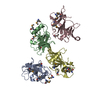
| ||||||||
|---|---|---|---|---|---|---|---|---|---|
| 1 | 
| ||||||||
| 2 | 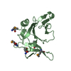
| ||||||||
| 3 | 
| ||||||||
| 4 | 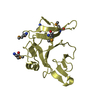
| ||||||||
| Unit cell |
|
-Components
| #1: Protein | Mass: 18308.777 Da / Num. of mol.: 4 / Fragment: S-type Pyocin domain residues 332-479 Source method: isolated from a genetically manipulated source Source: (gene. exp.) Erwinia carotovora (bacteria) / Strain: SCRI1043 / Gene: ECA1669 / Production host: #2: Water | ChemComp-HOH / |  Mass: 18.015 Da / Num. of mol.: 333 / Source method: isolated from a natural source / Formula: H2O Mass: 18.015 Da / Num. of mol.: 333 / Source method: isolated from a natural source / Formula: H2OHas protein modification | Y | |
|---|
-Experimental details
-Experiment
| Experiment | Method: X-RAY DIFFRACTION / Number of used crystals: 1 |
|---|
- Sample preparation
| Crystal | Density Matthews: 2.08 Å3/Da / Density % sol: 40.93 % |
|---|---|
| Crystal grow | Temperature: 277 K / Method: vapor diffusion, hanging drop / pH: 6.5 Details: Protein solution: 100mM NaCl, 5mM DTT, 0.02% NaN3, 10mM Tris-HCl (pH 7.5), Reservoir solution: 0.1M MES (pH 6.5), 30% PEG 5K MME, and 0.2M (NH4)2SO4, VAPOR DIFFUSION, HANGING DROP, temperature 277K |
-Data collection
| Diffraction | Mean temperature: 100 K |
|---|---|
| Diffraction source | Source: SYNCHROTRON / Site: NSLS  / Beamline: X4C / Wavelength: 0.97885 Å / Beamline: X4C / Wavelength: 0.97885 Å |
| Detector | Type: MAR CCD 165 mm / Detector: CCD / Date: Mar 25, 2010 / Details: mirrors |
| Radiation | Monochromator: Si 111 CHANNEL / Protocol: SINGLE WAVELENGTH / Monochromatic (M) / Laue (L): M / Scattering type: x-ray |
| Radiation wavelength | Wavelength: 0.97885 Å / Relative weight: 1 |
| Reflection | Resolution: 2.2→30 Å / Num. all: 60424 / Num. obs: 56618 / % possible obs: 93.7 % / Observed criterion σ(F): 0 / Observed criterion σ(I): 0 / Redundancy: 1.8 % / Biso Wilson estimate: 7.5 Å2 / Rmerge(I) obs: 0.048 / Rsym value: 0.046 / Net I/σ(I): 15.4 |
| Reflection shell | Resolution: 2.2→2.28 Å / Redundancy: 1.6 % / Rmerge(I) obs: 0.104 / Mean I/σ(I) obs: 7.3 / Num. unique all: 6084 / Rsym value: 0.121 / % possible all: 83.9 |
- Processing
| Software |
| ||||||||||||||||||||||||||||||||||||
|---|---|---|---|---|---|---|---|---|---|---|---|---|---|---|---|---|---|---|---|---|---|---|---|---|---|---|---|---|---|---|---|---|---|---|---|---|---|
| Refinement | Method to determine structure: SAD / Resolution: 2.2→19.41 Å / Rfactor Rfree error: 0.005 / Data cutoff high absF: 1000944.188 / Data cutoff low absF: 0 / Isotropic thermal model: RESTRAINED / Cross valid method: THROUGHOUT / σ(F): 2 / σ(I): 2 / Stereochemistry target values: Engh & Huber
| ||||||||||||||||||||||||||||||||||||
| Solvent computation | Solvent model: FLAT MODEL / Bsol: 36.021 Å2 / ksol: 0.4 e/Å3 | ||||||||||||||||||||||||||||||||||||
| Displacement parameters | Biso mean: 23.3 Å2
| ||||||||||||||||||||||||||||||||||||
| Refine analyze |
| ||||||||||||||||||||||||||||||||||||
| Refinement step | Cycle: LAST / Resolution: 2.2→19.41 Å
| ||||||||||||||||||||||||||||||||||||
| Refine LS restraints |
| ||||||||||||||||||||||||||||||||||||
| LS refinement shell | Resolution: 2.2→2.28 Å / Rfactor Rfree error: 0.029 / Total num. of bins used: 10
|
