Mass: 18.015 Da / Num. of mol.: 175 / Source method: isolated from a natural source / Formula: H2O
Has protein modification
Y
Sequence details
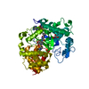
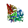
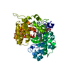
AUTHORS STATE THAT THEY BASED THE TEMPLATE FOR SMYD3 ON THE MGC CLONE (ACCESSION BC031010, VERSION ...AUTHORS STATE THAT THEY BASED THE TEMPLATE FOR SMYD3 ON THE MGC CLONE (ACCESSION BC031010, VERSION BC031010.1, GI:21410973)
-
Experimental details
-
Experiment
Experiment
Method: X-RAY DIFFRACTION / Number of used crystals: 1
-
Sample preparation
Crystal
Density Matthews: 2.17 Å3/Da / Density % sol: 43.25 %
Crystal grow
Temperature: 293 K / pH: 8 Details: 20% PEG3350, 0.2M Mg Formate, pH 8.0, VAPOR DIFFUSION, SITTING DROP, temperature 293K
Monochromator: DOUBLE-CRYSTAL MONOCHROMATOR / Protocol: SINGLE WAVELENGTH / Monochromatic (M) / Laue (L): M / Scattering type: x-ray
Radiation wavelength
Wavelength: 0.97935 Å / Relative weight: 1
Reflection
Resolution: 2.1→50 Å / Num. obs: 26234 / % possible obs: 100 % / Redundancy: 7.5 % / Rmerge(I) obs: 0.145 / Net I/σ(I): 6.4
Reflection shell
Resolution: 2.1→2.18 Å / Redundancy: 7.2 % / Rmerge(I) obs: 0.423 / % possible all: 100
-
Phasing
Phasing
Method: SAD
Phasing MAD
D res high: 2.09 Å / D res low: 44.7 Å / FOM acentric: 0.336 / FOM centric: 0.135 / Reflection acentric: 23070 / Reflection centric: 3102
Phasing MAD set
ID
R cullis acentric
R cullis centric
Highest resolution (Å)
Lowest resolution (Å)
Reflection acentric
Reflection centric
ISO_1
0
0
2.09
44.7
23070
3102
ANO_1
0.816
0
2.09
44.7
23064
0
Phasing MAD set shell
ID
Resolution (Å)
R cullis acentric
R cullis centric
Reflection acentric
Reflection centric
ISO_1
9.18-44.7
0
0
218
146
ISO_1
6.56-9.18
0
0
437
153
ISO_1
5.37-6.56
0
0
578
158
ISO_1
4.66-5.37
0
0
696
152
ISO_1
4.17-4.66
0
0
809
152
ISO_1
3.81-4.17
0
0
893
158
ISO_1
3.53-3.81
0
0
973
156
ISO_1
3.31-3.53
0
0
1062
155
ISO_1
3.12-3.31
0
0
1121
159
ISO_1
2.96-3.12
0
0
1203
155
ISO_1
2.82-2.96
0
0
1266
156
ISO_1
2.7-2.82
0
0
1316
159
ISO_1
2.6-2.7
0
0
1403
153
ISO_1
2.5-2.6
0
0
1430
159
ISO_1
2.42-2.5
0
0
1486
160
ISO_1
2.34-2.42
0
0
1536
145
ISO_1
2.27-2.34
0
0
1616
162
ISO_1
2.21-2.27
0
0
1657
153
ISO_1
2.15-2.21
0
0
1709
162
ISO_1
2.09-2.15
0
0
1661
149
ANO_1
9.18-44.7
0.448
0
218
0
ANO_1
6.56-9.18
0.392
0
437
0
ANO_1
5.37-6.56
0.424
0
578
0
ANO_1
4.66-5.37
0.564
0
695
0
ANO_1
4.17-4.66
0.656
0
809
0
ANO_1
3.81-4.17
0.694
0
892
0
ANO_1
3.53-3.81
0.697
0
972
0
ANO_1
3.31-3.53
0.681
0
1062
0
ANO_1
3.12-3.31
0.742
0
1121
0
ANO_1
2.96-3.12
0.71
0
1203
0
ANO_1
2.82-2.96
0.782
0
1266
0
ANO_1
2.7-2.82
0.786
0
1316
0
ANO_1
2.6-2.7
0.832
0
1403
0
ANO_1
2.5-2.6
0.878
0
1430
0
ANO_1
2.42-2.5
0.912
0
1486
0
ANO_1
2.34-2.42
0.931
0
1536
0
ANO_1
2.27-2.34
0.953
0
1616
0
ANO_1
2.21-2.27
0.965
0
1654
0
ANO_1
2.15-2.21
0.976
0
1709
0
ANO_1
2.09-2.15
0.984
0
1661
0
Phasing MAD set site
ID
Cartn x (Å)
Cartn y (Å)
Cartn z (Å)
Atom type symbol
B iso
Occupancy
1
-13.473
-14.43
-16.907
SE
21.99
1.86
2
-20.36
-1.728
-24.746
SE
24.57
1.48
3
-26.087
-15.522
-14.923
SE
23.95
1.45
4
-14.56
-16.6
-13.883
SE
28.68
1.58
5
-29.198
-1.78
-27.643
SE
27.34
1.29
6
-39.273
-4.852
-7.813
SE
41.82
1.6
7
-56.016
6.586
0.434
SE
32.62
1.14
8
-26.061
3.328
-21.465
SE
30.57
1.16
9
-23.109
-17.894
-21.765
SE
27.31
1.13
10
-16.98
-6.655
-14.42
SE
52.15
1.49
11
-34.763
-53.985
-1.295
SE
32.24
1
12
-44.591
-39.18
-11.785
SE
18.3
0.67
13
-39.774
-26.521
-23.767
SE
25.82
0.74
14
-41.404
-27.181
-3.562
SE
34.17
0.8
15
-44.664
-49.659
-4.14
SE
42.52
0.85
16
-26.099
-28.716
-16.638
SE
53.36
1.04
Phasing MAD shell
Resolution (Å)
FOM acentric
FOM centric
Reflection acentric
Reflection centric
9.18-44.7
0.63
0.211
218
146
6.56-9.18
0.645
0.202
437
153
5.37-6.56
0.614
0.211
578
158
4.66-5.37
0.541
0.144
696
152
4.17-4.66
0.459
0.12
809
152
3.81-4.17
0.439
0.142
893
158
3.53-3.81
0.448
0.11
973
156
3.31-3.53
0.438
0.129
1062
155
3.12-3.31
0.401
0.115
1121
159
2.96-3.12
0.417
0.131
1203
155
2.82-2.96
0.385
0.133
1266
156
2.7-2.82
0.383
0.14
1316
159
2.6-2.7
0.345
0.123
1403
153
2.5-2.6
0.304
0.132
1430
159
2.42-2.5
0.274
0.114
1486
160
2.34-2.42
0.255
0.114
1536
145
2.27-2.34
0.223
0.105
1616
162
2.21-2.27
0.203
0.119
1657
153
2.15-2.21
0.181
0.113
1709
162
2.09-2.15
0.169
0.104
1661
149
Phasing dm
Method: Solvent flattening and Histogram matching / Reflection: 26172
Phasing dm shell
Resolution (Å)
Delta phi final
FOM
Reflection
8.15-100
66.7
0.705
510
6.4-8.15
68.5
0.852
510
5.55-6.4
57.6
0.852
508
4.99-5.55
64.2
0.886
558
4.57-4.99
61.1
0.896
620
4.24-4.57
66.4
0.916
648
3.97-4.24
61.4
0.925
698
3.75-3.97
61.9
0.921
737
3.56-3.75
62.3
0.903
772
3.4-3.56
63.4
0.901
822
3.25-3.4
61.5
0.892
849
3.13-3.25
67.5
0.884
869
3.01-3.13
64.9
0.87
909
2.91-3.01
63.9
0.849
948
2.82-2.91
66.1
0.853
980
2.74-2.82
67
0.852
1014
2.66-2.74
66.1
0.844
1012
2.59-2.66
67.5
0.833
1066
2.53-2.59
69.9
0.843
1092
2.47-2.53
69.6
0.849
1104
2.41-2.47
73.8
0.829
1148
2.36-2.41
74.9
0.836
1150
2.31-2.36
74.6
0.824
1189
2.26-2.31
74.5
0.818
1201
2.22-2.26
76.4
0.81
1254
2.18-2.22
78.7
0.784
1240
2.14-2.18
79.6
0.783
1265
2.09-2.14
78.8
0.714
1499
-
Processing
Software
Name
Version
Classification
NB
DENZO
datareduction
SCALEPACK
datascaling
SHARP
phasing
DM
6.1
phasing
REFMAC
refmac_5.5.0109
refinement
PDB_EXTRACT
3.1
dataextraction
HKL-3000
datareduction
HKL-3000
datascaling
Refinement
Method to determine structure: SAD / Resolution: 2.1→44.7 Å / Cor.coef. Fo:Fc: 0.946 / Cor.coef. Fo:Fc free: 0.921 / Cross valid method: THROUGHOUT / ESU R: 0.215 / ESU R Free: 0.168 / Details: HYDROGENS HAVE BEEN ADDED IN THE RIDING POSITIONS
Rfactor
Num. reflection
% reflection
Selection details
Rfree
0.208
1328
5.074 %
RANDOM
Rwork
0.174
-
-
-
obs
0.17584
26172
99.7 %
-
Solvent computation
Ion probe radii: 0.8 Å / Shrinkage radii: 0.8 Å / VDW probe radii: 1.4 Å / Solvent model: BABINET MODEL PLUS MASK
Displacement parameters
Biso mean: 18.78 Å2
Baniso -1
Baniso -2
Baniso -3
1-
-0.027 Å2
0 Å2
0 Å2
2-
-
0.028 Å2
0 Å2
3-
-
-
-0.001 Å2
Refinement step
Cycle: LAST / Resolution: 2.1→44.7 Å
Protein
Nucleic acid
Ligand
Solvent
Total
Num. atoms
3339
0
30
175
3544
Refine LS restraints
Refine-ID
Type
Dev ideal
Dev ideal target
Number
X-RAY DIFFRACTION
r_bond_refined_d
0.011
0.022
3451
X-RAY DIFFRACTION
r_bond_other_d
X-RAY DIFFRACTION
r_angle_refined_deg
1.272
1.984
4666
X-RAY DIFFRACTION
r_angle_other_deg
X-RAY DIFFRACTION
r_dihedral_angle_1_deg
5.34
5
430
X-RAY DIFFRACTION
r_dihedral_angle_2_deg
33.04
24.013
157
X-RAY DIFFRACTION
r_dihedral_angle_3_deg
13.559
15
626
X-RAY DIFFRACTION
r_dihedral_angle_4_deg
17.047
15
26
X-RAY DIFFRACTION
r_chiral_restr
0.087
0.2
519
X-RAY DIFFRACTION
r_gen_planes_refined
0.005
0.021
2585
X-RAY DIFFRACTION
r_gen_planes_other
X-RAY DIFFRACTION
r_nbd_refined
0.195
0.2
1567
X-RAY DIFFRACTION
r_nbd_other
X-RAY DIFFRACTION
r_nbtor_refined
0.298
0.2
2402
X-RAY DIFFRACTION
r_nbtor_other
X-RAY DIFFRACTION
r_xyhbond_nbd_refined
0.107
0.2
195
X-RAY DIFFRACTION
r_xyhbond_nbd_other
X-RAY DIFFRACTION
r_metal_ion_refined
X-RAY DIFFRACTION
r_metal_ion_other
X-RAY DIFFRACTION
r_symmetry_vdw_refined
0.166
0.2
35
X-RAY DIFFRACTION
r_symmetry_vdw_other
X-RAY DIFFRACTION
r_symmetry_hbond_refined
0.126
0.2
12
X-RAY DIFFRACTION
r_symmetry_hbond_other
X-RAY DIFFRACTION
r_symmetry_metal_ion_refined
X-RAY DIFFRACTION
r_symmetry_metal_ion_other
X-RAY DIFFRACTION
r_mcbond_it
0.628
1.5
2129
X-RAY DIFFRACTION
r_mcbond_other
X-RAY DIFFRACTION
r_mcangle_it
1.261
2
3428
X-RAY DIFFRACTION
r_scbond_it
2.321
3
1322
X-RAY DIFFRACTION
r_scangle_it
3.853
4.5
1234
X-RAY DIFFRACTION
r_rigid_bond_restr
X-RAY DIFFRACTION
r_sphericity_free
X-RAY DIFFRACTION
r_sphericity_bonded
LS refinement shell
Resolution: 2.1→2.15 Å / Total num. of bins used: 20
Rfactor
Num. reflection
% reflection
Rfree
0.248
94
-
Rwork
0.187
1716
-
obs
-
-
96.02 %
Refinement TLS params.
Method: refined / Origin x: 12.208 Å / Origin y: 32.592 Å / Origin z: 12.902 Å
11
12
13
21
22
23
31
32
33
T
0.0082 Å2
-0.0053 Å2
-0.0015 Å2
-
0.0092 Å2
0.0014 Å2
-
-
0.0184 Å2
L
0.1889 °2
-0.1144 °2
-0.1562 °2
-
0.3384 °2
0.1611 °2
-
-
0.4733 °2
S
0.0155 Å °
0.0002 Å °
0.0098 Å °
-0.0274 Å °
-0.0026 Å °
-0.0124 Å °
-0.0542 Å °
0.0211 Å °
-0.0129 Å °
+
About Yorodumi
-
News
-
Feb 9, 2022. New format data for meta-information of EMDB entries
New format data for meta-information of EMDB entries
Version 3 of the EMDB header file is now the official format.
The previous official version 1.9 will be removed from the archive.
In the structure databanks used in Yorodumi, some data are registered as the other names, "COVID-19 virus" and "2019-nCoV". Here are the details of the virus and the list of structure data.
Jan 31, 2019. EMDB accession codes are about to change! (news from PDBe EMDB page)
EMDB accession codes are about to change! (news from PDBe EMDB page)
The allocation of 4 digits for EMDB accession codes will soon come to an end. Whilst these codes will remain in use, new EMDB accession codes will include an additional digit and will expand incrementally as the available range of codes is exhausted. The current 4-digit format prefixed with “EMD-” (i.e. EMD-XXXX) will advance to a 5-digit format (i.e. EMD-XXXXX), and so on. It is currently estimated that the 4-digit codes will be depleted around Spring 2019, at which point the 5-digit format will come into force.
The EM Navigator/Yorodumi systems omit the EMD- prefix.
Related info.:Q: What is EMD? / ID/Accession-code notation in Yorodumi/EM Navigator
Yorodumi is a browser for structure data from EMDB, PDB, SASBDB, etc.
This page is also the successor to EM Navigator detail page, and also detail information page/front-end page for Omokage search.
The word "yorodu" (or yorozu) is an old Japanese word meaning "ten thousand". "mi" (miru) is to see.
Related info.:EMDB / PDB / SASBDB / Comparison of 3 databanks / Yorodumi Search / Aug 31, 2016. New EM Navigator & Yorodumi / Yorodumi Papers / Jmol/JSmol / Function and homology information / Changes in new EM Navigator and Yorodumi
 Movie
Movie Controller

 Yorodumi
 Open data
 Basic information
 Components
 Keywords
 Function and homology information
 Homo sapiens (human)
 X-RAY DIFFRACTION /
  Authors
 Citation
 Structure visualization
 Downloads & links
 Other downloads


 PDBj
PDBj

 Assembly


 Mass: 65.409 Da / Num. of mol.: 3 / Source method: obtained synthetically / Formula: Zn
 Mass: 398.437 Da / Num. of mol.: 1 / Source method: obtained synthetically / Formula: C15H22N6O5S
 Mass: 18.015 Da / Num. of mol.: 175 / Source method: isolated from a natural source / Formula: H2O
 Sample preparation
 / Beamline: 19-ID / Wavelength: 0.97935
  Processing