Mass: 18.015 Da / Num. of mol.: 153 / Source method: isolated from a natural source / Formula: H2O
Sequence details
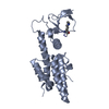
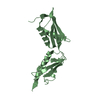
THE PROTEIN USED FOR CRYSTALLIZATION CONTAINED AMINO-ACIDS AS GIVEN IN DBREF. THIS PROTEIN LIKELY ...THE PROTEIN USED FOR CRYSTALLIZATION CONTAINED AMINO-ACIDS AS GIVEN IN DBREF. THIS PROTEIN LIKELY DEGRADED TO A TRUNCATED SPECIES DURING THE CRYSTALLIZATION EXPERIMENT. THEREFORE, CALCULATED VM AND VS VALUES ARE BASED ON AN ESTIMATE. THE AUTHORS ASSUMED RESIDUES 161 TO 281 TO BE PRESENT.
-
Experimental details
-
Experiment
Experiment
Method: X-RAY DIFFRACTION / Number of used crystals: 1
-
Sample preparation
Crystal
Density Matthews: 2.06 Å3/Da / Density % sol: 40.41 %
Crystal grow
Temperature: 292 K / Method: vapor diffusion, sitting drop / pH: 8 Details: 0.2 M calcium chloride, 20 % PEG3350, pH 8.0, VAPOR DIFFUSION, SITTING DROP, temperature 292K
Type: ADSC QUANTUM 315r / Detector: CCD / Date: Jun 14, 2009 / Details: SINGLE SILICON (111) MONOCHROMATOR
Radiation
Monochromator: SINGLE SILICON (111) / Protocol: SINGLE WAVELENGTH / Monochromatic (M) / Laue (L): M / Scattering type: x-ray
Radiation wavelength
Wavelength: 0.933 Å / Relative weight: 1
Reflection
Resolution: 2.1→50 Å / Num. obs: 23906 / % possible obs: 100 % / Observed criterion σ(I): -4 / Redundancy: 7.8 % / Biso Wilson estimate: 38.4 Å2 / Rmerge(I) obs: 0.102 / Net I/σ(I): 14.7
Reflection shell
Resolution: 2.1→2.13 Å / Redundancy: 7.7 % / Rmerge(I) obs: 0.498 / Mean I/σ(I) obs: 3.2 / % possible all: 100
-
Processing
Software
Name
Version
Classification
DNA
datacollection
PHASER
phasing
REFMAC
5.5.0102
refinement
HKL-2000
datareduction
SCALEPACK
datascaling
Refinement
Method to determine structure: MOLECULAR REPLACEMENT / Resolution: 2.2→50 Å / Cor.coef. Fo:Fc: 0.951 / Cor.coef. Fo:Fc free: 0.928 / SU B: 12.606 / SU ML: 0.145 / Cross valid method: THROUGHOUT / ESU R: 0.239 / ESU R Free: 0.202 / Stereochemistry target values: MAXIMUM LIKELIHOOD Details: HYDROGENS HAVE BEEN ADDED IN THE RIDING POSITIONS THE PROTEIN USED FOR CRYSTALLIZATION CONTAINED AMINO-ACIDS AS GIVEN IN DBREF. THIS PROTEIN LIKELY DEGRADED TO A TRUNCATED SPECIES DURING THE ...Details: HYDROGENS HAVE BEEN ADDED IN THE RIDING POSITIONS THE PROTEIN USED FOR CRYSTALLIZATION CONTAINED AMINO-ACIDS AS GIVEN IN DBREF. THIS PROTEIN LIKELY DEGRADED TO A TRUNCATED SPECIES DURING THE CRYSTALLIZATION EXPERIMENT. THEREFORE, CALCULATED VM AND VS VALUES ARE BASED ON AN ESTIMATE. THE AUTHORS ASSUMED RESIDUES 161 TO 281 TO BE PRESENT.
Rfactor
Num. reflection
% reflection
Selection details
Rfree
0.245
1056
5.1 %
RANDOM
Rwork
0.195
-
-
-
obs
0.198
19600
100 %
-
Solvent computation
Ion probe radii: 0.8 Å / Shrinkage radii: 0.8 Å / VDW probe radii: 1.4 Å / Solvent model: BABINET MODEL WITH MASK
Displacement parameters
Biso mean: 26.22 Å2
Baniso -1
Baniso -2
Baniso -3
1-
-1.05 Å2
-0.52 Å2
0 Å2
2-
-
-1.05 Å2
0 Å2
3-
-
-
1.57 Å2
Refinement step
Cycle: LAST / Resolution: 2.2→50 Å
Protein
Nucleic acid
Ligand
Solvent
Total
Num. atoms
2511
0
0
153
2664
Refine LS restraints
Refine-ID
Type
Dev ideal
Dev ideal target
Number
X-RAY DIFFRACTION
r_bond_refined_d
0.011
0.022
2550
X-RAY DIFFRACTION
r_bond_other_d
0.001
0.02
1695
X-RAY DIFFRACTION
r_angle_refined_deg
1.377
1.975
3480
X-RAY DIFFRACTION
r_angle_other_deg
1.11
3
4174
X-RAY DIFFRACTION
r_dihedral_angle_1_deg
6.045
5
323
X-RAY DIFFRACTION
r_dihedral_angle_2_deg
41.883
25.5
120
X-RAY DIFFRACTION
r_dihedral_angle_3_deg
17.972
15
422
X-RAY DIFFRACTION
r_dihedral_angle_4_deg
23.133
15
18
X-RAY DIFFRACTION
r_chiral_restr
0.076
0.2
417
X-RAY DIFFRACTION
r_gen_planes_refined
0.006
0.021
2844
X-RAY DIFFRACTION
r_gen_planes_other
0.001
0.02
454
X-RAY DIFFRACTION
r_nbd_refined
X-RAY DIFFRACTION
r_nbd_other
X-RAY DIFFRACTION
r_nbtor_refined
X-RAY DIFFRACTION
r_nbtor_other
X-RAY DIFFRACTION
r_xyhbond_nbd_refined
X-RAY DIFFRACTION
r_xyhbond_nbd_other
X-RAY DIFFRACTION
r_metal_ion_refined
X-RAY DIFFRACTION
r_metal_ion_other
X-RAY DIFFRACTION
r_symmetry_vdw_refined
X-RAY DIFFRACTION
r_symmetry_vdw_other
X-RAY DIFFRACTION
r_symmetry_hbond_refined
X-RAY DIFFRACTION
r_symmetry_hbond_other
X-RAY DIFFRACTION
r_symmetry_metal_ion_refined
X-RAY DIFFRACTION
r_symmetry_metal_ion_other
X-RAY DIFFRACTION
r_mcbond_it
0.583
1.5
1628
X-RAY DIFFRACTION
r_mcbond_other
0.098
1.5
649
X-RAY DIFFRACTION
r_mcangle_it
1.144
2
2668
X-RAY DIFFRACTION
r_scbond_it
1.843
3
922
X-RAY DIFFRACTION
r_scangle_it
3.333
4.5
812
X-RAY DIFFRACTION
r_rigid_bond_restr
X-RAY DIFFRACTION
r_sphericity_free
X-RAY DIFFRACTION
r_sphericity_bonded
LS refinement shell
Resolution: 2.2→2.26 Å / Total num. of bins used: 20
Rfactor
Num. reflection
% reflection
Rfree
0.28
80
-
Rwork
0.224
1477
-
obs
-
-
100 %
Refinement TLS params.
Method: refined / Refine-ID: X-RAY DIFFRACTION
ID
L11 (°2)
L12 (°2)
L13 (°2)
L22 (°2)
L23 (°2)
L33 (°2)
S11 (Å °)
S12 (Å °)
S13 (Å °)
S21 (Å °)
S22 (Å °)
S23 (Å °)
S31 (Å °)
S32 (Å °)
S33 (Å °)
T11 (Å2)
T12 (Å2)
T13 (Å2)
T22 (Å2)
T23 (Å2)
T33 (Å2)
Origin x (Å)
Origin y (Å)
Origin z (Å)
1
2.4022
1.2821
0.5893
2.2406
0.5647
0.8989
-0.0364
0.0254
-0.0708
-0.0384
0.0334
-0.1557
-0.0124
-0.0192
0.003
0.0252
-0.0111
-0.0045
0.0062
-0.0004
0.1451
47.9538
15.0815
-7.7063
2
5.6481
0.9166
-1.8956
1.424
-0.3479
2.2828
-0.2539
0.1554
-0.1847
0.0232
0.2267
-0.2443
0.1608
-0.1392
0.0272
0.075
-0.0127
-0.015
0.0762
-0.0525
0.0545
25.1852
-5.7911
-13.0622
3
2.9732
0.2944
1.1581
1.2008
-0.2295
1.1911
0.0472
0.0409
-0.1803
-0.0418
0.0164
-0.1469
-0.0676
-0.0657
-0.0637
0.1253
0.0015
0.0158
0.0762
-0.0109
0.0288
17.1824
-3.3259
7.7313
4
3.6807
1.632
-0.5717
3.3769
-1.9582
1.8789
-0.054
-0.1307
-0.0992
-0.0884
-0.1493
-0.1879
-0.001
0.1167
0.2033
0.0529
-0.0135
-0.0494
0.0181
0.0228
0.0814
47.4184
5.8392
12.5838
Refinement TLS group
ID
Refine-ID
Refine TLS-ID
Auth asym-ID
Auth seq-ID
1
X-RAY DIFFRACTION
1
A
217 - 296
2
X-RAY DIFFRACTION
2
A
297 - 381
3
X-RAY DIFFRACTION
3
B
217 - 296
4
X-RAY DIFFRACTION
4
B
297 - 381
+
About Yorodumi
-
News
-
Feb 9, 2022. New format data for meta-information of EMDB entries
New format data for meta-information of EMDB entries
Version 3 of the EMDB header file is now the official format.
The previous official version 1.9 will be removed from the archive.
In the structure databanks used in Yorodumi, some data are registered as the other names, "COVID-19 virus" and "2019-nCoV". Here are the details of the virus and the list of structure data.
Jan 31, 2019. EMDB accession codes are about to change! (news from PDBe EMDB page)
EMDB accession codes are about to change! (news from PDBe EMDB page)
The allocation of 4 digits for EMDB accession codes will soon come to an end. Whilst these codes will remain in use, new EMDB accession codes will include an additional digit and will expand incrementally as the available range of codes is exhausted. The current 4-digit format prefixed with “EMD-” (i.e. EMD-XXXX) will advance to a 5-digit format (i.e. EMD-XXXXX), and so on. It is currently estimated that the 4-digit codes will be depleted around Spring 2019, at which point the 5-digit format will come into force.
The EM Navigator/Yorodumi systems omit the EMD- prefix.
Related info.:Q: What is EMD? / ID/Accession-code notation in Yorodumi/EM Navigator
Yorodumi is a browser for structure data from EMDB, PDB, SASBDB, etc.
This page is also the successor to EM Navigator detail page, and also detail information page/front-end page for Omokage search.
The word "yorodu" (or yorozu) is an old Japanese word meaning "ten thousand". "mi" (miru) is to see.
Related info.:EMDB / PDB / SASBDB / Comparison of 3 databanks / Yorodumi Search / Aug 31, 2016. New EM Navigator & Yorodumi / Yorodumi Papers / Jmol/JSmol / Function and homology information / Changes in new EM Navigator and Yorodumi
 Movie
Movie Controller


 Open data
 Basic information
 Components
 Keywords
 Function and homology information
 Nostoc sp. (bacteria)
 X-RAY DIFFRACTION /
  Authors
 Citation
 Structure visualization
 Downloads & links
 Other downloads


 PDBj
PDBj Assembly

 Mass: 18.015 Da / Num. of mol.: 153 / Source method: isolated from a natural source / Formula: H2O
 Sample preparation
 / Beamline: ID23-1 / Wavelength: 0.933
  Processing