| 登録情報 | データベース: PDB / ID: 3maz
|
|---|
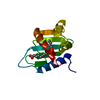
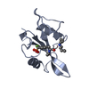
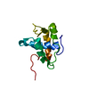
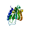
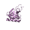
| タイトル | Crystal Structure of the Human BRDG1/STAP-1 SH2 Domain in Complex with the NTAL pTyr136 Peptide |
|---|
 要素 要素 | - CheD family protein
- Signal-transducing adaptor protein 1
|
|---|
 キーワード キーワード | SIGNALING PROTEIN / modular domain / phosphotyrosine / specificity / Cytoplasm / Phosphoprotein / SH2 domain |
|---|
| 機能・相同性 |  機能・相同性情報 機能・相同性情報
negative regulation of microglial cell migration / negative regulation of ruffle assembly / macrophage colony-stimulating factor receptor binding / positive regulation of B cell receptor signaling pathway / negative regulation of macrophage colony-stimulating factor signaling pathway / positive regulation of microglial cell mediated cytotoxicity / positive regulation of microglial cell activation / positive regulation of phagocytosis, engulfment / negative regulation of macrophage chemotaxis / transmembrane receptor protein tyrosine kinase adaptor activity ...negative regulation of microglial cell migration / negative regulation of ruffle assembly / macrophage colony-stimulating factor receptor binding / positive regulation of B cell receptor signaling pathway / negative regulation of macrophage colony-stimulating factor signaling pathway / positive regulation of microglial cell mediated cytotoxicity / positive regulation of microglial cell activation / positive regulation of phagocytosis, engulfment / negative regulation of macrophage chemotaxis / transmembrane receptor protein tyrosine kinase adaptor activity / mast cell degranulation / B cell activation / protein tyrosine kinase activator activity / Role of LAT2/NTAL/LAB on calcium mobilization / signaling adaptor activity / phosphotyrosine residue binding / SH2 domain binding / cell surface receptor protein tyrosine kinase signaling pathway / B cell receptor signaling pathway / calcium-mediated signaling / phospholipid binding / cellular response to lipopolysaccharide / adaptive immune response / intracellular signal transduction / nuclear body / membrane raft / positive regulation of gene expression / protein kinase binding / protein-containing complex / mitochondrion / extracellular exosome / nucleus / plasma membrane / cytoplasm / cytosol類似検索 - 分子機能 Linker for activation of T-cells family member 2 / Linker for activation of T-cells family member 2 / STAP1, SH2 domain / Signal-transducing adaptor protein STAP1/STAP2 / SH2 domain / SHC Adaptor Protein / PH domain / PH domain profile. / Pleckstrin homology domain. / Pleckstrin homology domain ...Linker for activation of T-cells family member 2 / Linker for activation of T-cells family member 2 / STAP1, SH2 domain / Signal-transducing adaptor protein STAP1/STAP2 / SH2 domain / SHC Adaptor Protein / PH domain / PH domain profile. / Pleckstrin homology domain. / Pleckstrin homology domain / SH2 domain / Src homology 2 (SH2) domain profile. / Src homology 2 domains / SH2 domain / SH2 domain superfamily / PH-like domain superfamily / 2-Layer Sandwich / Alpha Beta類似検索 - ドメイン・相同性 MALONATE ION / Linker for activation of T-cells family member 2 / Signal-transducing adaptor protein 1類似検索 - 構成要素 |
|---|
| 生物種 |  Homo sapiens (ヒト) Homo sapiens (ヒト) |
|---|
| 手法 |  X線回折 / 分子置換 / 解像度: 1.9 Å X線回折 / 分子置換 / 解像度: 1.9 Å |
|---|
 データ登録者 データ登録者 | Kaneko, T. / Huang, H. / Zhao, B. / Li, L. / Liu, H. / Voss, C.K. / Wu, C. / Schiller, M.R. / Li, S.S. |
|---|
 引用 引用 | ジャーナル: Sci.Signal. / 年: 2010
タイトル: Loops govern SH2 domain specificity by controlling access to binding pockets.
著者: Kaneko, T. / Huang, H. / Zhao, B. / Li, L. / Liu, H. / Voss, C.K. / Wu, C. / Schiller, M.R. / Li, S.S. |
|---|
| 履歴 | | 登録 | 2010年3月24日 | 登録サイト: RCSB / 処理サイト: RCSB |
|---|
| 改定 1.0 | 2010年5月12日 | Provider: repository / タイプ: Initial release |
|---|
| 改定 1.1 | 2011年7月13日 | Group: Version format compliance |
|---|
| 改定 1.2 | 2021年10月6日 | Group: Database references / Derived calculations
カテゴリ: database_2 / struct_conn ...database_2 / struct_conn / struct_ref_seq_dif / struct_site
Item: _database_2.pdbx_DOI / _database_2.pdbx_database_accession ..._database_2.pdbx_DOI / _database_2.pdbx_database_accession / _struct_conn.pdbx_leaving_atom_flag / _struct_ref_seq_dif.details / _struct_site.pdbx_auth_asym_id / _struct_site.pdbx_auth_comp_id / _struct_site.pdbx_auth_seq_id |
|---|
| 改定 1.3 | 2023年9月6日 | Group: Data collection / Refinement description
カテゴリ: chem_comp_atom / chem_comp_bond / pdbx_initial_refinement_model |
|---|
| 改定 1.4 | 2023年11月22日 | Group: Data collection / カテゴリ: chem_comp_atom / chem_comp_bond / Item: _chem_comp_atom.atom_id / _chem_comp_bond.atom_id_2 |
|---|
| 改定 1.5 | 2024年11月20日 | Group: Structure summary
カテゴリ: pdbx_entry_details / pdbx_modification_feature |
|---|
|
|---|
 ムービー
ムービー コントローラー


 データを開く
 基本情報
基本情報 構造の表示
構造の表示 ダウンロードとリンク
 その他のダウンロード
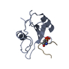


 PDBj
PDBj


 集合体
集合体


 分子量: 102.046 Da / 分子数: 1 / 由来タイプ: 合成 / 式: C3H2O4
 分子量: 18.015 Da / 分子数: 83 / 由来タイプ: 天然 / 式: H2O
 試料調製
試料調製 解析
解析