 Movie
Movie Controller

[English] 日本語
 Yorodumi
- PDB-3m7a: Crystal structure of Saro_0823 (YP_496102.1) a protein of unknown... -
+ Open data

- Basic information
| Entry | Database: PDB / ID: 3m7a | ||||||
|---|---|---|---|---|---|---|---|
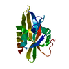
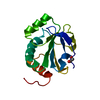
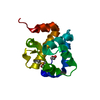
| Title | Crystal structure of Saro_0823 (YP_496102.1) a protein of unknown function from NOVOSPHINGOBIUM AROMATICIVORANS DSM 12444 at 1.22 A resolution | ||||||
 Components Components | uncharacterized protein | ||||||
 Keywords Keywords | structural genomics / unknown function / Joint Center for Structural Genomics / JCSG / Protein Structure Initiative / PSI-2 / Uncharacterized ACR / COG1430 | ||||||
| Function / homology | Protein of unknown function DUF192 / Protein of unknown function DUF192 / Saro_0823-like superfamily / Uncharacterized ACR, COG1430 / Prokaryotic membrane lipoprotein lipid attachment site profile. / Jelly Rolls / Sandwich / Mainly Beta / DUF192 domain-containing protein Function and homology information Function and homology information | ||||||
| Biological species |  Novosphingobium aromaticivorans (bacteria) Novosphingobium aromaticivorans (bacteria) | ||||||
| Method |  X-RAY DIFFRACTION / SYNCHROTRON / MAD / Resolution: 1.22 Å X-RAY DIFFRACTION / SYNCHROTRON / MAD / Resolution: 1.22 Å | ||||||
 Authors Authors | Joint Center for Structural Genomics (JCSG) | ||||||
 Citation Citation | Journal: To be Published Title: Crystal structure of Saro_0823 (YP_496102.1) a protein of unknown function from NOVOSPHINGOBIUM AROMATICIVORANS DSM 12444 at 1.22 A resolution Authors: Joint Center for Structural Genomics (JCSG) | ||||||
| History |
|
- Structure visualization
| Structure viewer | Molecule: MolmilJmol/JSmol |
|---|
- Downloads & links
-Download
| PDBx/mmCIF format | 3m7a.cif.gz | 144.9 KB | Display | PDBx/mmCIF format |
|---|---|---|---|---|
| PDB format | pdb3m7a.ent.gz | 114 KB | Display | PDB format |
| PDBx/mmJSON format | 3m7a.json.gz | Tree view | PDBx/mmJSON format | |
| Others |  Other downloads Other downloads |
-Validation report
| Summary document | 3m7a_validation.pdf.gz | 440.2 KB | Display | wwPDB validaton report |
|---|---|---|---|---|
| Full document | 3m7a_full_validation.pdf.gz | 441.3 KB | Display | |
| Data in XML | 3m7a_validation.xml.gz | 17.5 KB | Display | |
| Data in CIF | 3m7a_validation.cif.gz | 27.4 KB | Display | |
| Arichive directory | https://data.pdbj.org/pub/pdb/validation_reports/m7/3m7aftp://data.pdbj.org/pub/pdb/validation_reports/m7/3m7a | HTTPS FTP |
-Related structure data
| Similar structure data | |
|---|---|
| Other databases |

-Links
 PDBj
PDBj


- Assembly
| Deposited unit | 
| ||||||||
|---|---|---|---|---|---|---|---|---|---|
| 1 | 
| ||||||||
| 2 | 
| ||||||||
| Unit cell |
| ||||||||
| Components on special symmetry positions |
|
-Components
| #1: Protein | Mass: 15126.579 Da / Num. of mol.: 2 Source method: isolated from a genetically manipulated source Source: (gene. exp.) Novosphingobium aromaticivorans (bacteria)Strain: DSM 12444 / Gene: Saro_0823 / Plasmid: SpeedET / Production host: #2: Chemical | ChemComp-EDO /   Mass: 62.068 Da / Num. of mol.: 6 / Source method: obtained synthetically / Formula: C2H6O2 Mass: 62.068 Da / Num. of mol.: 6 / Source method: obtained synthetically / Formula: C2H6O2#3: Water | ChemComp-HOH / |  Mass: 18.015 Da / Num. of mol.: 458 / Source method: isolated from a natural source / Formula: H2O Mass: 18.015 Da / Num. of mol.: 458 / Source method: isolated from a natural source / Formula: H2OHas protein modification | Y | Sequence details | THIS CONSTRUCT (RESIDUES 27-165) WAS EXPRESSED WITH AN N-TERMINAL PURIFICATION TAG ...THIS CONSTRUCT (RESIDUES 27-165) WAS EXPRESSED WITH AN N-TERMINAL PURIFICATI | |
|---|
-Experimental details
-Experiment
| Experiment | Method: X-RAY DIFFRACTION / Number of used crystals: 1 |
|---|
- Sample preparation
| Crystal | Density Matthews: 2.05 Å3/Da / Density % sol: 39.91 % |
|---|---|
| Crystal grow | Temperature: 277 K / Method: vapor diffusion, sitting drop / pH: 8.86 Details: 29.5000% polyethylene glycol 4000, 0.2000M magnesium chloride, 0.1M TRIS pH 8.86, NANODROP', VAPOR DIFFUSION, SITTING DROP, temperature 277K |
-Data collection
| Diffraction | Mean temperature: 100 K | ||||||||||||||||||||||||||||||||||||||||||||||||||||||||||||||||||
|---|---|---|---|---|---|---|---|---|---|---|---|---|---|---|---|---|---|---|---|---|---|---|---|---|---|---|---|---|---|---|---|---|---|---|---|---|---|---|---|---|---|---|---|---|---|---|---|---|---|---|---|---|---|---|---|---|---|---|---|---|---|---|---|---|---|---|---|
| Diffraction source | Source: SYNCHROTRON / Site: SSRL  / Beamline: BL11-1 / Wavelength: 0.97883,0.91837 / Beamline: BL11-1 / Wavelength: 0.97883,0.91837 | ||||||||||||||||||||||||||||||||||||||||||||||||||||||||||||||||||
| Detector | Type: MARMOSAIC 325 mm CCD / Detector: CCD / Date: Dec 2, 2009 / Details: Flat mirror (vertical focusing) | ||||||||||||||||||||||||||||||||||||||||||||||||||||||||||||||||||
| Radiation | Monochromator: Single crystal Si(111) bent monochromator (horizontal focusing) Protocol: MAD / Monochromatic (M) / Laue (L): M / Scattering type: x-ray | ||||||||||||||||||||||||||||||||||||||||||||||||||||||||||||||||||
| Radiation wavelength |
| ||||||||||||||||||||||||||||||||||||||||||||||||||||||||||||||||||
| Reflection | Resolution: 1.22→45.531 Å / Num. obs: 71683 / % possible obs: 98.6 % / Observed criterion σ(I): -3 / Biso Wilson estimate: 8.037 Å2 / Rmerge(I) obs: 0.09 / Net I/σ(I): 10.24 | ||||||||||||||||||||||||||||||||||||||||||||||||||||||||||||||||||
| Reflection shell |
|
-Phasing
| Phasing | Method: MAD |
|---|
- Processing
| Software |
| ||||||||||||||||||||||||||||||||||||||||||||||||||||||||||||||||||||||||||||||||||||||||||
|---|---|---|---|---|---|---|---|---|---|---|---|---|---|---|---|---|---|---|---|---|---|---|---|---|---|---|---|---|---|---|---|---|---|---|---|---|---|---|---|---|---|---|---|---|---|---|---|---|---|---|---|---|---|---|---|---|---|---|---|---|---|---|---|---|---|---|---|---|---|---|---|---|---|---|---|---|---|---|---|---|---|---|---|---|---|---|---|---|---|---|---|
| Refinement | Method to determine structure: MAD / Resolution: 1.22→45.531 Å / Cor.coef. Fo:Fc: 0.982 / Cor.coef. Fo:Fc free: 0.976 / Occupancy max: 1 / Occupancy min: 0.2 / SU B: 1.216 / SU ML: 0.024 / Cross valid method: THROUGHOUT / σ(F): 0 / ESU R: 0.037 / ESU R Free: 0.037 Stereochemistry target values: MAXIMUM LIKELIHOOD WITH PHASES Details: 1. HYDROGENS HAVE BEEN ADDED IN THE RIDING POSITIONS. 2. A MET-INHIBITION PROTOCOL WAS USED FOR SELENOMETHIONINE INCORPORATION DURING PROTEIN EXPRESSION. THE OCCUPANCY OF THE SE ATOMS IN THE ...Details: 1. HYDROGENS HAVE BEEN ADDED IN THE RIDING POSITIONS. 2. A MET-INHIBITION PROTOCOL WAS USED FOR SELENOMETHIONINE INCORPORATION DURING PROTEIN EXPRESSION. THE OCCUPANCY OF THE SE ATOMS IN THE MSE RESIDUES WAS REDUCED TO 0.75 FOR THE REDUCED SCATTERING POWER DUE TO PARTIAL S-MET INCORPORATION. 3. ETHYLENE GLYCOL (EDO) MODELED WERE PRESENT IN CRYO CONDITIONS.
| ||||||||||||||||||||||||||||||||||||||||||||||||||||||||||||||||||||||||||||||||||||||||||
| Solvent computation | Ion probe radii: 0.8 Å / Shrinkage radii: 0.8 Å / VDW probe radii: 1.2 Å / Solvent model: MASK | ||||||||||||||||||||||||||||||||||||||||||||||||||||||||||||||||||||||||||||||||||||||||||
| Displacement parameters | Biso max: 70.62 Å2 / Biso mean: 11.968 Å2 / Biso min: 3.61 Å2
| ||||||||||||||||||||||||||||||||||||||||||||||||||||||||||||||||||||||||||||||||||||||||||
| Refinement step | Cycle: LAST / Resolution: 1.22→45.531 Å
| ||||||||||||||||||||||||||||||||||||||||||||||||||||||||||||||||||||||||||||||||||||||||||
| Refine LS restraints |
| ||||||||||||||||||||||||||||||||||||||||||||||||||||||||||||||||||||||||||||||||||||||||||
| LS refinement shell | Resolution: 1.22→1.252 Å / Total num. of bins used: 20
|
