 Movie
Movie Controller

[English] 日本語
 Yorodumi
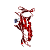
- PDB-3m0n: Plasmodium vivax 6-pyruvoyltetrahydropterin synthase (PTPS), E37A... -
+ Open data

- Basic information
| Entry | Database: PDB / ID: 3m0n | ||||||
|---|---|---|---|---|---|---|---|
| Title | Plasmodium vivax 6-pyruvoyltetrahydropterin synthase (PTPS), E37A catalytic residue mutant | ||||||
 Components Components | Putative 6-pyruvoyl tetrahydrobiopterin synthase | ||||||
 Keywords Keywords | BIOSYNTHETIC PROTEIN / PTS / PTP SYNTHASE / PTPS / METAL-BINDING / TETRAHYDROBIOPTERIN BIOSYNTHESIS / FOLATE BIOSYNTHESIS / Structural Genomics / Medical Structural Genomics of Pathogenic Protozoa / MSGPP | ||||||
| Function / homology |  Function and homology information Function and homology information6-pyruvoyltetrahydropterin synthase / 6-pyruvoyltetrahydropterin synthase activity / tetrahydrobiopterin biosynthetic process / metal ion binding Similarity search - Function | ||||||
| Biological species |  | ||||||
| Method |  X-RAY DIFFRACTION / SYNCHROTRON / FOURIER SYNTHESIS / Resolution: 1.9 Å X-RAY DIFFRACTION / SYNCHROTRON / FOURIER SYNTHESIS / Resolution: 1.9 Å | ||||||
 Authors Authors | Larson, E.T. / Merritt, E.A. / Medical Structural Genomics of Pathogenic Protozoa (MSGPP) | ||||||
 Citation Citation | Journal: to be published Title: Structural analysis of the dual-functional 6-pyruvoyltetrahydropterin synthase from Malaria parasites. Authors: Larson, E.T. / Bosch, J. / Kim, J.E. / Kelley, A. / Castaneda, L. / Napuli, A. / Mueller, N. / Verlinde, C.L.M.J. / Van Voorhis, W.C. / Buckner, F.S. / Fan, E. / Hol, W.G.J. / Merritt, E.A. | ||||||
| History |
|
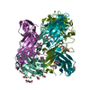
- Structure visualization
| Structure viewer | Molecule: MolmilJmol/JSmol |
|---|
- Downloads & links
-Download
| PDBx/mmCIF format | 3m0n.cif.gz | 87.7 KB | Display | PDBx/mmCIF format |
|---|---|---|---|---|
| PDB format | pdb3m0n.ent.gz | 66 KB | Display | PDB format |
| PDBx/mmJSON format | 3m0n.json.gz | Tree view | PDBx/mmJSON format | |
| Others |  Other downloads Other downloads |
-Validation report
| Arichive directory | https://data.pdbj.org/pub/pdb/validation_reports/m0/3m0nftp://data.pdbj.org/pub/pdb/validation_reports/m0/3m0n | HTTPS FTP |
|---|
-Related structure data
| Related structure data | 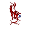 3lx3SC  3lzeC S: Starting model for refinement C: citing same article ( |
|---|---|
| Similar structure data | |
| Other databases |


-Links
 PDBj
PDBj
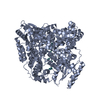
- Assembly
| Deposited unit | 
| ||||||||
|---|---|---|---|---|---|---|---|---|---|
| 1 | x 6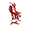
| ||||||||
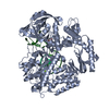
| Unit cell |
|
-Components
| #1: Protein | Mass: 20934.863 Da / Num. of mol.: 1 / Mutation: E37A Source method: isolated from a genetically manipulated source Details: E37A mutation confirmed from plasmid DNA sequence Source: (gene. exp.) Gene: PVX_114505 / Plasmid: BG1861 / Production host:  References: UniProt: A5K2B2, 6-pyruvoyltetrahydropterin synthase |
|---|---|
| #2: Chemical | ChemComp-ZN /   Mass: 65.409 Da / Num. of mol.: 1 / Source method: obtained synthetically / Formula: Zn Mass: 65.409 Da / Num. of mol.: 1 / Source method: obtained synthetically / Formula: Zn |
| #3: Chemical | ChemComp-PE0 /   Mass: 163.137 Da / Num. of mol.: 1 / Source method: obtained synthetically / Formula: C6H5N5O Mass: 163.137 Da / Num. of mol.: 1 / Source method: obtained synthetically / Formula: C6H5N5O |
| #4: Water | ChemComp-HOH /  Mass: 18.015 Da / Num. of mol.: 59 / Source method: isolated from a natural source / Formula: H2O Mass: 18.015 Da / Num. of mol.: 59 / Source method: isolated from a natural source / Formula: H2O |
| Sequence details | E37A MUTATION CONFIRMED FROM PLASMID DNA SEQUENCE |
-Experimental details
-Experiment
| Experiment | Method: X-RAY DIFFRACTION / Number of used crystals: 1 |
|---|
- Sample preparation
| Crystal | Density Matthews: 2.96 Å3/Da / Density % sol: 58.47 % |
|---|---|
| Crystal grow | Temperature: 298 K / Method: vapor diffusion, hanging drop / pH: 5.2 Details: 2 ul 25 mg/ml protein (in SGPP buffer) mixed with 2 ul 0.1 M sodium acetate (pH 5.2), 28% PEG 3350, 5 mM DTT; cryoprotected by 5 sec dip in 96 mM sodium acetate (pH 5.2), 24% PEG 3350, 134 ...Details: 2 ul 25 mg/ml protein (in SGPP buffer) mixed with 2 ul 0.1 M sodium acetate (pH 5.2), 28% PEG 3350, 5 mM DTT; cryoprotected by 5 sec dip in 96 mM sodium acetate (pH 5.2), 24% PEG 3350, 134 mM NaCl, 20% glycerol, vapor diffusion, hanging drop, temperature 298K |
-Data collection
| Diffraction | Mean temperature: 100 K |
|---|---|
| Diffraction source | Source: SYNCHROTRON / Site: SSRL  / Beamline: BL9-2 / Wavelength: 0.9795 Å / Beamline: BL9-2 / Wavelength: 0.9795 Å |
| Detector | Type: MARMOSAIC 325 mm CCD / Detector: CCD / Date: Feb 4, 2010 |
| Radiation | Monochromator: crystal / Protocol: SINGLE WAVELENGTH / Monochromatic (M) / Laue (L): M / Scattering type: x-ray |
| Radiation wavelength | Wavelength: 0.9795 Å / Relative weight: 1 |
| Reflection | Resolution: 1.9→50 Å / Num. obs: 19474 / % possible obs: 99.8 % / Observed criterion σ(I): 5 / Redundancy: 9.3 % / Biso Wilson estimate: 43 Å2 / Rmerge(I) obs: 0.079 / Χ2: 1.064 / Net I/σ(I): 12.9 |
| Reflection shell | Resolution: 1.9→1.97 Å / Redundancy: 9.4 % / Rmerge(I) obs: 0.683 / Mean I/σ(I) obs: 2.4 / Num. unique all: 1947 / Χ2: 0.98 / % possible all: 100 |
- Processing
| Software |
| |||||||||||||||||||||||||||||||||||||||||||||||||||||||||||||||||||||||||||||||||||||||||||||||||||||||||||||||||||||||||||||||||||||||||||||||||||||||||||||||||||||||||||||||||||||||||||||||||||||||||||||||||||||||||||||||||
|---|---|---|---|---|---|---|---|---|---|---|---|---|---|---|---|---|---|---|---|---|---|---|---|---|---|---|---|---|---|---|---|---|---|---|---|---|---|---|---|---|---|---|---|---|---|---|---|---|---|---|---|---|---|---|---|---|---|---|---|---|---|---|---|---|---|---|---|---|---|---|---|---|---|---|---|---|---|---|---|---|---|---|---|---|---|---|---|---|---|---|---|---|---|---|---|---|---|---|---|---|---|---|---|---|---|---|---|---|---|---|---|---|---|---|---|---|---|---|---|---|---|---|---|---|---|---|---|---|---|---|---|---|---|---|---|---|---|---|---|---|---|---|---|---|---|---|---|---|---|---|---|---|---|---|---|---|---|---|---|---|---|---|---|---|---|---|---|---|---|---|---|---|---|---|---|---|---|---|---|---|---|---|---|---|---|---|---|---|---|---|---|---|---|---|---|---|---|---|---|---|---|---|---|---|---|---|---|---|---|---|---|---|---|---|---|---|---|---|---|---|---|---|---|---|---|---|
| Refinement | Method to determine structure: FOURIER SYNTHESIS Starting model: 3lx3, ligand removed and with E37 mutated to A Resolution: 1.9→35.3 Å / Cor.coef. Fo:Fc: 0.96 / Cor.coef. Fo:Fc free: 0.947 / Occupancy max: 1 / Occupancy min: 0.4 / SU B: 6.565 / SU ML: 0.089 / Cross valid method: THROUGHOUT / σ(F): 0 / ESU R: 0.128 / ESU R Free: 0.122 / Stereochemistry target values: MAXIMUM LIKELIHOOD Details: HYDROGENS HAVE BEEN ADDED IN THE RIDING POSITIONS; U VALUES: WITH TLS ADDED. Identity of ligand in active site unknown but clear density for pterine ring of substrate or product present so modeled as such
| |||||||||||||||||||||||||||||||||||||||||||||||||||||||||||||||||||||||||||||||||||||||||||||||||||||||||||||||||||||||||||||||||||||||||||||||||||||||||||||||||||||||||||||||||||||||||||||||||||||||||||||||||||||||||||||||||
| Solvent computation | Ion probe radii: 0.8 Å / Shrinkage radii: 0.8 Å / VDW probe radii: 1.4 Å / Solvent model: BABINET MODEL WITH MASK | |||||||||||||||||||||||||||||||||||||||||||||||||||||||||||||||||||||||||||||||||||||||||||||||||||||||||||||||||||||||||||||||||||||||||||||||||||||||||||||||||||||||||||||||||||||||||||||||||||||||||||||||||||||||||||||||||
| Displacement parameters | Biso max: 108.34 Å2 / Biso mean: 49.913 Å2 / Biso min: 23.57 Å2
| |||||||||||||||||||||||||||||||||||||||||||||||||||||||||||||||||||||||||||||||||||||||||||||||||||||||||||||||||||||||||||||||||||||||||||||||||||||||||||||||||||||||||||||||||||||||||||||||||||||||||||||||||||||||||||||||||
| Refinement step | Cycle: LAST / Resolution: 1.9→35.3 Å
| |||||||||||||||||||||||||||||||||||||||||||||||||||||||||||||||||||||||||||||||||||||||||||||||||||||||||||||||||||||||||||||||||||||||||||||||||||||||||||||||||||||||||||||||||||||||||||||||||||||||||||||||||||||||||||||||||
| Refine LS restraints |
| |||||||||||||||||||||||||||||||||||||||||||||||||||||||||||||||||||||||||||||||||||||||||||||||||||||||||||||||||||||||||||||||||||||||||||||||||||||||||||||||||||||||||||||||||||||||||||||||||||||||||||||||||||||||||||||||||
| LS refinement shell | Resolution: 1.9→1.95 Å / Total num. of bins used: 20
| |||||||||||||||||||||||||||||||||||||||||||||||||||||||||||||||||||||||||||||||||||||||||||||||||||||||||||||||||||||||||||||||||||||||||||||||||||||||||||||||||||||||||||||||||||||||||||||||||||||||||||||||||||||||||||||||||
| Refinement TLS params. | Method: refined / Refine-ID: X-RAY DIFFRACTION
| |||||||||||||||||||||||||||||||||||||||||||||||||||||||||||||||||||||||||||||||||||||||||||||||||||||||||||||||||||||||||||||||||||||||||||||||||||||||||||||||||||||||||||||||||||||||||||||||||||||||||||||||||||||||||||||||||
| Refinement TLS group |
|
