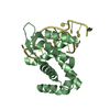
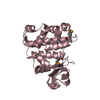
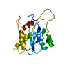
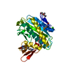
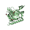
- PDB-3lwx: Crystal structure of Na(+)-translocating NADH-quinone reductase s... -
+
Open data
ID or keywords:
Loading...
-
Basic information
Entry
Database: PDB / ID: 3lwx
Title
Crystal structure of Na(+)-translocating NADH-quinone reductase subunit C (YP_001302508.1) from Parabacteroides distasonis ATCC 8503 at 1.10 A resolution
Components
NADH:ubiquinone oxidoreductase, Na translocating, C subunit
Keywords
OXIDOREDUCTASE / Na(+)-translocating NADH-quinone reductase subunit C / Structural Genomics / Joint Center for Structural Genomics / JCSG / Protein Structure Initiative / PSI-2 / Transport / Ubiquinone
Function / homology
Function and homology information
NADH:ubiquinone reductase (Na+-transporting) / oxidoreductase activity, acting on NAD(P)H, quinone or similar compound as acceptor / sodium ion transport / FMN binding / iron ion binding / plasma membrane Similarity search - Function
Mass: 18.015 Da / Num. of mol.: 370 / Source method: isolated from a natural source / Formula: H2O
Has protein modification
Y
Sequence details
THE CONSTRUCT WAS EXPRESSED WITH A PURIFICATION TAG MGSDKIHHHHHHENLYFQG. THE TAG WAS REMOVED WITH ...THE CONSTRUCT WAS EXPRESSED WITH A PURIFICATION TAG MGSDKIHHHHHHENLYFQG. THE TAG WAS REMOVED WITH TEV PROTEASE LEAVING ONLY A GLYCINE (0) FOLLOWED BY THE TARGET SEQUENCE. THE CLONED CONSTRUCT CONTAINS RESIDUES 73-270 OF THE FULL LENGTH PROTEIN.
-
Experimental details
-
Experiment
Experiment
Method: X-RAY DIFFRACTION / Number of used crystals: 1
-
Sample preparation
Crystal
Density Matthews: 2.15 Å3/Da / Density % sol: 42.92 %
Crystal grow
Temperature: 277 K / Method: vapor diffusion, sitting drop / pH: 8.5 Details: 15.0000% Glycerol, 0.1700M NaOAc, 25.5000% PEG-4000, 0.1M TRIS pH 8.5, NANODROP, VAPOR DIFFUSION, SITTING DROP, temperature 277K
Monochromator: Double crystal monochromator / Protocol: SINGLE WAVELENGTH / Monochromatic (M) / Laue (L): M / Scattering type: x-ray
Radiation wavelength
Wavelength: 0.97861 Å / Relative weight: 1
Reflection
Resolution: 1.1→29.739 Å / Num. obs: 74185 / % possible obs: 95.2 % / Observed criterion σ(I): -3 / Biso Wilson estimate: 7.951 Å2 / Rmerge(I) obs: 0.041 / Net I/σ(I): 14.69
Reflection shell
Resolution (Å)
Rmerge(I) obs
Mean I/σ(I) obs
Num. measured obs
Num. unique obs
% possible all
1.1-1.14
0.534
2.2
33755
13515
89.9
1.14-1.18
0.405
2.8
30531
12058
92.6
1.18-1.24
0.342
3.3
39522
15520
93.4
1.24-1.3
0.268
4.3
32994
12933
94.6
1.3-1.39
0.194
5.7
39554
15425
94.8
1.39-1.49
0.135
7.8
33871
13211
95.7
1.49-1.64
0.08
11.8
36907
14340
96.4
1.64-1.88
0.054
18.6
44179
14632
97.3
1.88-2.37
0.032
35.5
62672
14612
98.4
2.37-29.739
0.025
50.9
64769
14688
99.1
-
Phasing
Phasing
Method: SAD
-
Processing
Software
Name
Version
Classification
NB
REFMAC
5.5.0102
refinement
PHENIX
refinement
SHELX
phasing
MolProbity
3beta29
modelbuilding
XSCALE
datascaling
PDB_EXTRACT
3.006
dataextraction
XDS
datareduction
SHELXD
phasing
autoSHARP
phasing
Refinement
Method to determine structure: SAD / Resolution: 1.1→29.739 Å / Cor.coef. Fo:Fc: 0.984 / Cor.coef. Fo:Fc free: 0.978 / Occupancy max: 1 / Occupancy min: 0.2 / SU B: 0.806 / SU ML: 0.017 / Cross valid method: THROUGHOUT / σ(F): 0 / ESU R: 0.028 / ESU R Free: 0.028 Stereochemistry target values: MAXIMUM LIKELIHOOD WITH PHASES Details: 1.HYDROGENS HAVE BEEN ADDED IN THE RIDING POSITIONS. 2. A MET-INHIBITION PROTOCOL WAS USED FOR SELENOMETHIONINE INCORPORATION DURING PROTEIN EXPRESSION. THE OCCUPANCY OF THE SE ATOMS IN THE ...Details: 1.HYDROGENS HAVE BEEN ADDED IN THE RIDING POSITIONS. 2. A MET-INHIBITION PROTOCOL WAS USED FOR SELENOMETHIONINE INCORPORATION DURING PROTEIN EXPRESSION. THE OCCUPANCY OF THE SE ATOMS IN THE MSE RESIDUES WAS REDUCED TO 0.75 TO ACCOUNT FOR THE REDUCED SCATTERING POWER DUE TO PARTIAL S-MET INCORPORATION. 3. GLYCEROL (GOL) FROM THE CRYSTALLIZATION SOLUTION WERE MODELED INTO THE STRUCTURE.
Rfactor
Num. reflection
% reflection
Selection details
Rfree
0.138
3700
5 %
RANDOM
Rwork
0.116
-
-
-
obs
0.117
74143
96 %
-
Solvent computation
Ion probe radii: 0.8 Å / Shrinkage radii: 0.8 Å / VDW probe radii: 1.4 Å / Solvent model: BABINET MODEL WITH MASK
In the structure databanks used in Yorodumi, some data are registered as the other names, "COVID-19 virus" and "2019-nCoV". Here are the details of the virus and the list of structure data.
Jan 31, 2019. EMDB accession codes are about to change! (news from PDBe EMDB page)
EMDB accession codes are about to change! (news from PDBe EMDB page)
The allocation of 4 digits for EMDB accession codes will soon come to an end. Whilst these codes will remain in use, new EMDB accession codes will include an additional digit and will expand incrementally as the available range of codes is exhausted. The current 4-digit format prefixed with “EMD-” (i.e. EMD-XXXX) will advance to a 5-digit format (i.e. EMD-XXXXX), and so on. It is currently estimated that the 4-digit codes will be depleted around Spring 2019, at which point the 5-digit format will come into force.
The EM Navigator/Yorodumi systems omit the EMD- prefix.
Related info.:Q: What is EMD? / ID/Accession-code notation in Yorodumi/EM Navigator
Yorodumi is a browser for structure data from EMDB, PDB, SASBDB, etc.
This page is also the successor to EM Navigator detail page, and also detail information page/front-end page for Omokage search.
The word "yorodu" (or yorozu) is an old Japanese word meaning "ten thousand". "mi" (miru) is to see.
Related info.:EMDB / PDB / SASBDB / Comparison of 3 databanks / Yorodumi Search / Aug 31, 2016. New EM Navigator & Yorodumi / Yorodumi Papers / Jmol/JSmol / Function and homology information / Changes in new EM Navigator and Yorodumi
 Movie
Movie Controller

 Yorodumi
 Open data
 Basic information
 Components
 Keywords
 Function and homology information
 Parabacteroides distasonis (bacteria)
 X-RAY DIFFRACTION /
  Authors
 Citation
 Structure visualization
 Downloads & links
 Other downloads
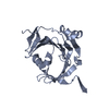


 PDBj
PDBj
 Assembly
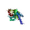

 Mass: 92.094 Da / Num. of mol.: 3 / Source method: obtained synthetically / Formula: C3H8O3
 Mass: 18.015 Da / Num. of mol.: 370 / Source method: isolated from a natural source / Formula: H2O
 Sample preparation
 / Beamline: BL9-2 / Wavelength: 0.97861
  Processing