- PDB-3ln3: Crystal structure of Putative reductase (NP_038806.2) from MUS MU... -
+
Open data
ID or keywords:
Loading...
-
Basic information
Entry
Database: PDB / ID: 3ln3
Title
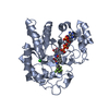
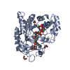
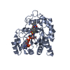
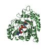
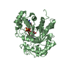
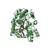
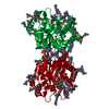
Crystal structure of Putative reductase (NP_038806.2) from MUS MUSCULUS at 1.18 A resolution
Components
Dihydrodiol dehydrogenase
Keywords
OXIDOREDUCTASE / Putative reductase / Structural Genomics / Joint Center for Structural Genomics / JCSG / Protein Structure Initiative / PSI-2
Function / homology
Function and homology information
Synthesis of bile acids and bile salts via 24-hydroxycholesterol / chlordecone reductase activity / RA biosynthesis pathway / Retinoid metabolism and transport / trans-1,2-dihydrobenzene-1,2-diol dehydrogenase activity / Synthesis of bile acids and bile salts via 7alpha-hydroxycholesterol / Synthesis of bile acids and bile salts via 27-hydroxycholesterol / Synthesis of Prostaglandins (PG) and Thromboxanes (TX) / Prednisone ADME / prostaglandin D2 11-ketoreductase activity ...Synthesis of bile acids and bile salts via 24-hydroxycholesterol / chlordecone reductase activity / RA biosynthesis pathway / Retinoid metabolism and transport / trans-1,2-dihydrobenzene-1,2-diol dehydrogenase activity / Synthesis of bile acids and bile salts via 7alpha-hydroxycholesterol / Synthesis of bile acids and bile salts via 27-hydroxycholesterol / Synthesis of Prostaglandins (PG) and Thromboxanes (TX) / Prednisone ADME / prostaglandin D2 11-ketoreductase activity / ketoreductase activity / 15-hydroxyprostaglandin-D dehydrogenase (NADP+) activity / Delta4-3-oxosteroid 5beta-reductase activity / : / testosterone dehydrogenase (NADP+) activity / 3-alpha-hydroxysteroid 3-dehydrogenase [NAD(P)+] activity / androsterone dehydrogenase [NAD(P)+] activity / ketosteroid monooxygenase activity / progesterone metabolic process / carboxylic acid binding / all-trans-retinol dehydrogenase (NAD+) activity / alcohol dehydrogenase (NADP+) activity / prostaglandin H2 endoperoxidase reductase activity / Oxidoreductases; Acting on the CH-OH group of donors; With NAD+ or NADP+ as acceptor / bile acid binding / daunorubicin metabolic process / doxorubicin metabolic process / retinal dehydrogenase (NAD+) activity / aldose reductase (NADPH) activity / oxidoreductase activity, acting on NAD(P)H, quinone or similar compound as acceptor / prostaglandin metabolic process / xenobiotic metabolic process / oxidoreductase activity / endoplasmic reticulum / nucleus / cytoplasm / cytosol Similarity search - Function
Aldo-keto reductase family 1 member C / Aldo/keto reductase family signature 1. / NADP-dependent oxidoreductase domain / Aldo/keto reductase family signature 2. / Aldo/keto reductase, conserved site / Aldo-keto reductase / NADP-dependent oxidoreductase domain / Aldo/keto reductase family / NADP-dependent oxidoreductase domain superfamily / TIM Barrel ...Aldo-keto reductase family 1 member C / Aldo/keto reductase family signature 1. / NADP-dependent oxidoreductase domain / Aldo/keto reductase family signature 2. / Aldo/keto reductase, conserved site / Aldo-keto reductase / NADP-dependent oxidoreductase domain / Aldo/keto reductase family / NADP-dependent oxidoreductase domain superfamily / TIM Barrel / Alpha-Beta Barrel / Alpha Beta Similarity search - Domain/homology
NICOTINAMIDE-ADENINE-DINUCLEOTIDE / Dihydrodiol dehydrogenase / Aldo-keto reductase family 1 member C13 Similarity search - Component
Biological species
Mus musculus (house mouse)
Method
X-RAY DIFFRACTION / SYNCHROTRON / SAD / Resolution: 1.18 Å
Mass: 18.015 Da / Num. of mol.: 395 / Source method: isolated from a natural source / Formula: H2O
Has protein modification
Y
Sequence details
1.THIS CONSTRUCT WAS EXPRESSED WITH A PURIFICATION TAG MGSDKIHHHHHHENLYFQG. THE TAG WAS REMOVED ...1.THIS CONSTRUCT WAS EXPRESSED WITH A PURIFICATION TAG MGSDKIHHHHHHENLYFQG. THE TAG WAS REMOVED WITH TEV PROTEASE LEAVING ONLY A GLYCINE (0) FOLLOWED BY THE TARGET SEQUENCE. 2.THE PROTEIN WAS REDUCTIVELY METHYLATED PRIOR TO CRYSTALLIZATION.
-
Experimental details
-
Experiment
Experiment
Method: X-RAY DIFFRACTION / Number of used crystals: 1
-
Sample preparation
Crystal
Density Matthews: 2.29 Å3/Da / Density % sol: 46.17 %
Crystal grow
Temperature: 293 K / Method: vapor diffusion, sitting drop / pH: 8 Details: 2.4000M ammonium sulfate, 0.1M TRIS pH 8.0, NANODROP, VAPOR DIFFUSION, SITTING DROP, temperature 293K
Monochromator: Double crystal monochromator / Protocol: SINGLE WAVELENGTH / Monochromatic (M) / Laue (L): M / Scattering type: x-ray
Radiation wavelength
Wavelength: 0.97931 Å / Relative weight: 1
Reflection
Resolution: 1.18→28.895 Å / Num. obs: 110769 / % possible obs: 95 % / Observed criterion σ(I): -3 / Biso Wilson estimate: 7.922 Å2 / Rmerge(I) obs: 0.064 / Net I/σ(I): 6.23
Reflection shell
Resolution (Å)
Rmerge(I) obs
Mean I/σ(I) obs
Num. measured obs
Num. unique obs
% possible all
1.18-1.22
0.457
1.5
21167
14203
67.4
1.22-1.27
0.403
1.7
33225
21719
94.6
1.27-1.33
0.328
2.1
34483
22419
97.4
1.33-1.4
0.264
2.6
33706
21780
98.4
1.4-1.49
0.197
3.4
35070
22528
99.1
1.49-1.6
0.136
4.7
33041
21071
99.3
1.6-1.76
0.1
6.3
34882
22060
99.1
1.76-2.02
0.066
9.1
35674
22393
98.7
2.02-2.54
0.046
13
34836
21583
98.2
2.54-28.895
0.037
16.6
35475
21504
96.2
-
Phasing
Phasing
Method: SAD
-
Processing
Software
Name
Version
Classification
NB
REFMAC
5.5.0053
refinement
PHENIX
refinement
SHELX
phasing
MolProbity
3beta29
modelbuilding
XSCALE
datascaling
PDB_EXTRACT
3.006
dataextraction
XDS
datareduction
SHELXD
phasing
autoSHARP
phasing
Refinement
Method to determine structure: SAD / Resolution: 1.18→28.895 Å / Cor.coef. Fo:Fc: 0.978 / Cor.coef. Fo:Fc free: 0.973 / Occupancy max: 1 / Occupancy min: 0.25 / SU B: 1.046 / SU ML: 0.021 / Cross valid method: THROUGHOUT / σ(F): 0 / ESU R: 0.033 / ESU R Free: 0.033 Stereochemistry target values: MAXIMUM LIKELIHOOD WITH PHASES Details: HYDROGENS HAVE BEEN ADDED IN THE RIDING POSITIONS 1.HYDROGENS HAVE BEEN ADDED IN THE RIDING POSITIONS. 2.A MET-INHIBITION PROTOCOL WAS USED FOR SELENOMETHIONINE INCORPORATION DURING PROTEIN ...Details: HYDROGENS HAVE BEEN ADDED IN THE RIDING POSITIONS 1.HYDROGENS HAVE BEEN ADDED IN THE RIDING POSITIONS. 2.A MET-INHIBITION PROTOCOL WAS USED FOR SELENOMETHIONINE INCORPORATION DURING PROTEIN EXPRESSION. THE OCCUPANCY OF THE SE ATOMS IN THE MSE RESIDUES WAS REDUCED TO 0.75 FOR THE REDUCED SCATTERING POWER DUE TO PARTIAL S-MET INCORPORATION. 3.SULFATE (SO4) FROM THE CRYSTALLIZATION SOLUTION HAS BEEN MODELED IN THE SOLVENT STRUCTURE. 4.ENDOGENOUS NICOTINAMIDE ADENINE DINUCLEOTIDE (NAD), WHICH IS THE EXPECTED COFACTOR FOR THIS PROTEIN, HAS BEEN MODELED AT THE PUTATIVE ACTIVE SITE. THE NAD SHOWS SOME DISORDER IN THE STRUCTURE. 5.(4R)-2-METHYLPENTANE-2,4-DIOL (MRD), WHICH MAY BE A SUBSTRATE MIMIC, HAS BEEN MODELED IN THE PUTATIVE ACTIVE SITE BASED ON THE ELECTRON DENSITY. HOWEVER, IT IS POSSIBLE THAT THIS MAY BE SOME OTHER COMPOUND. 6.THE PROTEIN WAS SUBJECTED TO REDUCTIVE METHYLATION PRIOR TO CRYSTALLIZATION. LYSINE 84 APPEARS TO HAVE BEEN PROTECTED FROM REDUCTIVE METHYLATION AND WAS MODELED AS LYSINE. ALL OTHER LYSINES HAVE BEEN MODELED AS N-DIMETHYL-LYSINE (MLY).
Rfactor
Num. reflection
% reflection
Selection details
Rfree
0.156
5543
5 %
RANDOM
Rwork
0.131
-
-
-
obs
0.132
110769
97.84 %
-
Solvent computation
Ion probe radii: 0.8 Å / Shrinkage radii: 0.8 Å / VDW probe radii: 1.4 Å / Solvent model: BABINET MODEL WITH MASK
In the structure databanks used in Yorodumi, some data are registered as the other names, "COVID-19 virus" and "2019-nCoV". Here are the details of the virus and the list of structure data.
Jan 31, 2019. EMDB accession codes are about to change! (news from PDBe EMDB page)
EMDB accession codes are about to change! (news from PDBe EMDB page)
The allocation of 4 digits for EMDB accession codes will soon come to an end. Whilst these codes will remain in use, new EMDB accession codes will include an additional digit and will expand incrementally as the available range of codes is exhausted. The current 4-digit format prefixed with “EMD-” (i.e. EMD-XXXX) will advance to a 5-digit format (i.e. EMD-XXXXX), and so on. It is currently estimated that the 4-digit codes will be depleted around Spring 2019, at which point the 5-digit format will come into force.
The EM Navigator/Yorodumi systems omit the EMD- prefix.
Related info.:Q: What is EMD? / ID/Accession-code notation in Yorodumi/EM Navigator
Yorodumi is a browser for structure data from EMDB, PDB, SASBDB, etc.
This page is also the successor to EM Navigator detail page, and also detail information page/front-end page for Omokage search.
The word "yorodu" (or yorozu) is an old Japanese word meaning "ten thousand". "mi" (miru) is to see.
Related info.:EMDB / PDB / SASBDB / Comparison of 3 databanks / Yorodumi Search / Aug 31, 2016. New EM Navigator & Yorodumi / Yorodumi Papers / Jmol/JSmol / Function and homology information / Changes in new EM Navigator and Yorodumi
 Movie
Movie Controller

 Yorodumi
 Open data
 Basic information
 Components
 Keywords
 Function and homology information
 X-RAY DIFFRACTION /
  Authors
 Citation
 Structure visualization
 Downloads & links
 Other downloads


 PDBj
PDBj


 Assembly
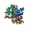


 Mass: 96.063 Da / Num. of mol.: 3 / Source method: obtained synthetically / Formula: SO4
 Mass: 663.425 Da / Num. of mol.: 1 / Source method: obtained synthetically / Formula: C21H27N7O14P2 / Comment: NAD*YM
 Mass: 118.174 Da / Num. of mol.: 1 / Source method: obtained synthetically / Formula: C6H14O2 / Comment: precipitant*YM
 Mass: 18.015 Da / Num. of mol.: 395 / Source method: isolated from a natural source / Formula: H2O
 Sample preparation
 / Beamline: BL9-2 / Wavelength: 0.97931
  Processing