 Movie
Movie Controller

[English] 日本語
 Yorodumi
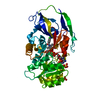
- PDB-3lmb: The crystal structure of the protein OLEI01261 with unknown funct... -
+ Open data

- Basic information
| Entry | Database: PDB / ID: 3lmb | ||||||
|---|---|---|---|---|---|---|---|
| Title | The crystal structure of the protein OLEI01261 with unknown function from Chlorobaculum tepidum TLS | ||||||
 Components Components | Uncharacterized protein | ||||||
 Keywords Keywords | Structural Genomics / Unknown function / protein OLEI01261 / Chlorobaculum tepidum TLS / PSI2 / MCSG / Protein Structure Initiative / Midwest Center for Structural Genomics | ||||||
| Function / homology | Hotdog Thioesterase / Thiol Ester Dehydrase; Chain A / Roll / Alpha Beta Function and homology information Function and homology information | ||||||
| Biological species |  Oleispira antarctica RB-8 (bacteria) Oleispira antarctica RB-8 (bacteria) | ||||||
| Method |  X-RAY DIFFRACTION / SYNCHROTRON / SAD / Resolution: 2.1 Å X-RAY DIFFRACTION / SYNCHROTRON / SAD / Resolution: 2.1 Å | ||||||
 Authors Authors | Zhang, R. / Evdokimova, E. / Egorova, O. / Savchenko, A. / Edwards, A. / Joachimiak, A. / Midwest Center for Structural Genomics (MCSG) | ||||||
 Citation Citation | Journal: Nat Commun / Year: 2013 Title: Genome sequence and functional genomic analysis of the oil-degrading bacterium Oleispira antarctica. Authors: Kube, M. / Chernikova, T.N. / Al-Ramahi, Y. / Beloqui, A. / Lopez-Cortez, N. / Guazzaroni, M.E. / Heipieper, H.J. / Klages, S. / Kotsyurbenko, O.R. / Langer, I. / Nechitaylo, T.Y. / ...Authors: Kube, M. / Chernikova, T.N. / Al-Ramahi, Y. / Beloqui, A. / Lopez-Cortez, N. / Guazzaroni, M.E. / Heipieper, H.J. / Klages, S. / Kotsyurbenko, O.R. / Langer, I. / Nechitaylo, T.Y. / Lunsdorf, H. / Fernandez, M. / Juarez, S. / Ciordia, S. / Singer, A. / Kagan, O. / Egorova, O. / Alain Petit, P. / Stogios, P. / Kim, Y. / Tchigvintsev, A. / Flick, R. / Denaro, R. / Genovese, M. / Albar, J.P. / Reva, O.N. / Martinez-Gomariz, M. / Tran, H. / Ferrer, M. / Savchenko, A. / Yakunin, A.F. / Yakimov, M.M. / Golyshina, O.V. / Reinhardt, R. / Golyshin, P.N. | ||||||
| History |
|
- Structure visualization
| Structure viewer | Molecule: MolmilJmol/JSmol |
|---|
- Downloads & links
-Download
| PDBx/mmCIF format | 3lmb.cif.gz | 79.8 KB | Display | PDBx/mmCIF format |
|---|---|---|---|---|
| PDB format | pdb3lmb.ent.gz | 61.1 KB | Display | PDB format |
| PDBx/mmJSON format | 3lmb.json.gz | Tree view | PDBx/mmJSON format | |
| Others |  Other downloads Other downloads |
-Validation report
| Arichive directory | https://data.pdbj.org/pub/pdb/validation_reports/lm/3lmbftp://data.pdbj.org/pub/pdb/validation_reports/lm/3lmb | HTTPS FTP |
|---|
-Related structure data
| Related structure data |  3i4qC 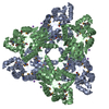 3iruC  3lnpC 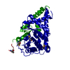 3m16C  3qvmC  3v77C  3vcrC C: citing same article ( |
|---|---|
| Similar structure data | |
| Other databases |

-Links
 PDBj
PDBj- Assembly
| Deposited unit | 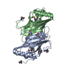
| ||||||||
|---|---|---|---|---|---|---|---|---|---|
| 1 |
| ||||||||
| Unit cell |
|
-Components
| #1: Protein | Mass: 18818.492 Da / Num. of mol.: 2 Source method: isolated from a genetically manipulated source Source: (gene. exp.) Oleispira antarctica RB-8 (bacteria) / Plasmid: pMCSG7 / Production host: #2: Water | ChemComp-HOH / |  Mass: 18.015 Da / Num. of mol.: 134 / Source method: isolated from a natural source / Formula: H2O Mass: 18.015 Da / Num. of mol.: 134 / Source method: isolated from a natural source / Formula: H2OHas protein modification | Y | |
|---|
-Experimental details
-Experiment
| Experiment | Method: X-RAY DIFFRACTION / Number of used crystals: 1 |
|---|
- Sample preparation
| Crystal | Density Matthews: 2.25 Å3/Da / Density % sol: 45.22 % |
|---|---|
| Crystal grow | Temperature: 289 K / Method: vapor diffusion, sitting drop Details: 0.2 M Mg Formate, 20% PEG3350, VAPOR DIFFUSION, SITTING DROP, temperature 289K |
-Data collection
| Diffraction | Mean temperature: 100 K |
|---|---|
| Diffraction source | Source: SYNCHROTRON / Site: APS  / Beamline: 19-ID / Wavelength: 0.9794 Å / Beamline: 19-ID / Wavelength: 0.9794 Å |
| Detector | Type: ADSC QUANTUM 315r / Detector: CCD / Date: Nov 11, 2009 / Details: mirrors |
| Radiation | Monochromator: Si 111 CHANNEL / Protocol: SINGLE WAVELENGTH / Monochromatic (M) / Laue (L): M / Scattering type: x-ray |
| Radiation wavelength | Wavelength: 0.9794 Å / Relative weight: 1 |
| Reflection | Resolution: 2.1→53.83 Å / Num. all: 19342 / Num. obs: 19329 / % possible obs: 99.93 % / Observed criterion σ(F): 2 / Observed criterion σ(I): 2 / Redundancy: 9.5 % / Rmerge(I) obs: 0.08 / Net I/σ(I): 26.43 |
| Reflection shell | Resolution: 2.1→2.154 Å / Redundancy: 9.4 % / Rmerge(I) obs: 0.416 / Mean I/σ(I) obs: 2.73 / Num. unique all: 1487 / % possible all: 99.8 |
- Processing
| Software |
| |||||||||||||||||||||||||||||||||||||||||||||||||||||||||||||||||
|---|---|---|---|---|---|---|---|---|---|---|---|---|---|---|---|---|---|---|---|---|---|---|---|---|---|---|---|---|---|---|---|---|---|---|---|---|---|---|---|---|---|---|---|---|---|---|---|---|---|---|---|---|---|---|---|---|---|---|---|---|---|---|---|---|---|---|
| Refinement | Method to determine structure: SAD / Resolution: 2.1→53.83 Å / Cor.coef. Fo:Fc: 0.961 / Cor.coef. Fo:Fc free: 0.936 / SU B: 9.102 / SU ML: 0.111 / TLS residual ADP flag: LIKELY RESIDUAL / Cross valid method: THROUGHOUT / ESU R: 0.203 / ESU R Free: 0.178 Stereochemistry target values: MAXIMUM LIKELIHOOD WITH PHASES Details: HYDROGENS HAVE BEEN ADDED IN THE RIDING POSITIONS
| |||||||||||||||||||||||||||||||||||||||||||||||||||||||||||||||||
| Solvent computation | Ion probe radii: 0.8 Å / Shrinkage radii: 0.8 Å / VDW probe radii: 1.4 Å / Solvent model: MASK | |||||||||||||||||||||||||||||||||||||||||||||||||||||||||||||||||
| Displacement parameters | Biso mean: 23.599 Å2
| |||||||||||||||||||||||||||||||||||||||||||||||||||||||||||||||||
| Refinement step | Cycle: LAST / Resolution: 2.1→53.83 Å
| |||||||||||||||||||||||||||||||||||||||||||||||||||||||||||||||||
| Refine LS restraints |
| |||||||||||||||||||||||||||||||||||||||||||||||||||||||||||||||||
| LS refinement shell | Resolution: 2.1→2.154 Å / Total num. of bins used: 20
| |||||||||||||||||||||||||||||||||||||||||||||||||||||||||||||||||
| Refinement TLS params. | Method: refined / Origin x: 10.714 Å / Origin y: 34.586 Å / Origin z: 0.184 Å
| |||||||||||||||||||||||||||||||||||||||||||||||||||||||||||||||||
| Refinement TLS group |
|
