 Movie
Movie Controller

[English] 日本語
 Yorodumi
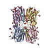
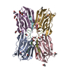
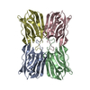
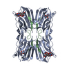
- PDB-3lm1: Crystal Structure Analysis of Maclura pomifera agglutinin complex... -
+ Open data

- Basic information
| Entry | Database: PDB / ID: 3lm1 | ||||||
|---|---|---|---|---|---|---|---|
| Title | Crystal Structure Analysis of Maclura pomifera agglutinin complex with p-nitrophenyl-GalNAc | ||||||
 Components Components |
| ||||||
 Keywords Keywords | SUGAR BINDING PROTEIN / Maclura pomifera agglutinin / MPA / MPA complex / (p)-nitrophenyl-GalNAc / Lectin | ||||||
| Function / homology |  Function and homology information Function and homology information | ||||||
| Biological species |  Maclura pomifera (Osage orange) Maclura pomifera (Osage orange) | ||||||
| Method |  X-RAY DIFFRACTION / SYNCHROTRON / MOLECULAR REPLACEMENT / Resolution: 2.1 Å X-RAY DIFFRACTION / SYNCHROTRON / MOLECULAR REPLACEMENT / Resolution: 2.1 Å | ||||||
 Authors Authors | Huang, J. / Xu, Z. / Wang, D. / Ogato, C. / Hirama, T. / Palczewski, K. / Hazen, S.L. / Lee, X. / Young, N.M. | ||||||
 Citation Citation | Journal: Glycobiology / Year: 2010 Title: Characterization of the secondary binding sites of Maclura pomifera agglutinin by glycan array and crystallographic analyses. Authors: Huang, J. / Xu, Z. / Wang, D. / Ogata, C.M. / Palczewski, K. / Lee, X. / Young, N.M. | ||||||
| History |
|
- Structure visualization
| Structure viewer | Molecule: MolmilJmol/JSmol |
|---|
- Downloads & links
-Download
| PDBx/mmCIF format | 3lm1.cif.gz | 258.1 KB | Display | PDBx/mmCIF format |
|---|---|---|---|---|
| PDB format | pdb3lm1.ent.gz | 210.2 KB | Display | PDB format |
| PDBx/mmJSON format | 3lm1.json.gz | Tree view | PDBx/mmJSON format | |
| Others |  Other downloads Other downloads |
-Validation report
| Arichive directory | https://data.pdbj.org/pub/pdb/validation_reports/lm/3lm1ftp://data.pdbj.org/pub/pdb/validation_reports/lm/3lm1 | HTTPS FTP |
|---|
-Related structure data
| Related structure data | 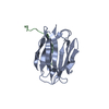 3llyC 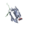 3llzC 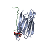 1jotS C: citing same article ( S: Starting model for refinement |
|---|---|
| Similar structure data |
-Links
 PDBj
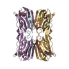
PDBj- Assembly
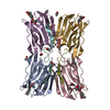
| Deposited unit | 
| ||||||||
|---|---|---|---|---|---|---|---|---|---|
| 1 | 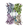
| ||||||||
| 2 | 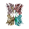
| ||||||||
| Unit cell |
| ||||||||
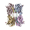
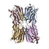
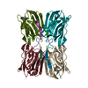
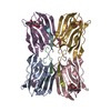
| Details | In one asymmetric unit, there are two agglutinin tetramers |
-Components
| #1: Protein | Mass: 14768.595 Da / Num. of mol.: 8 / Source method: isolated from a natural source / Details: From the seeds of the Moraceae plant family / Source: (natural) Maclura pomifera (Osage orange) / References: UniProt: P18674#2: Protein/peptide | Mass: 1615.790 Da / Num. of mol.: 8 / Source method: isolated from a natural source / Details: From the seeds of the Moraceae plant family / Source: (natural) Maclura pomifera (Osage orange) / References: UniProt: P18676#3: Sugar | ChemComp-LEC / 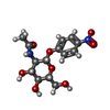  Type: D-saccharide / Mass: 342.301 Da / Num. of mol.: 7 / Source method: obtained synthetically / Formula: C14H18N2O8 Type: D-saccharide / Mass: 342.301 Da / Num. of mol.: 7 / Source method: obtained synthetically / Formula: C14H18N2O8#4: Water | ChemComp-HOH / |  Mass: 18.015 Da / Num. of mol.: 879 / Source method: isolated from a natural source / Formula: H2O Mass: 18.015 Da / Num. of mol.: 879 / Source method: isolated from a natural source / Formula: H2O |
|---|
-Experimental details
-Experiment
| Experiment | Method: X-RAY DIFFRACTION / Number of used crystals: 1 |
|---|
- Sample preparation
| Crystal | Density Matthews: 3.53 Å3/Da / Density % sol: 65.18 % |
|---|---|
| Crystal grow | Temperature: 298 K / Method: vapor diffusion, sitting drop / pH: 7 Details: 0.5M of lithium sulfate, 12% PEG 8000, 1% octyl-beta-D-glucopyranoside, 0.1M Hepes, pH 7.0 in the reservoir solution. The sitting drop is made by protein (28mg/mL) and equal volumn of ...Details: 0.5M of lithium sulfate, 12% PEG 8000, 1% octyl-beta-D-glucopyranoside, 0.1M Hepes, pH 7.0 in the reservoir solution. The sitting drop is made by protein (28mg/mL) and equal volumn of reservoir solution in the presence of p-nitrophenyl-alpha-GalNAc., VAPOR DIFFUSION, SITTING DROP, temperature 298K |
-Data collection
| Diffraction | Mean temperature: 100 K |
|---|---|
| Diffraction source | Source: SYNCHROTRON / Site: NSLS  / Beamline: X4A / Wavelength: 0.9793 Å / Beamline: X4A / Wavelength: 0.9793 Å |
| Detector | Type: ADSC QUANTUM 4r / Detector: CCD / Date: Jun 30, 2000 / Details: mirrors |
| Radiation | Monochromator: KOHZU double crystal monochromator with a sagittally focused second crystal Protocol: SINGLE WAVELENGTH / Monochromatic (M) / Laue (L): M / Scattering type: x-ray |
| Radiation wavelength | Wavelength: 0.9793 Å / Relative weight: 1 |
| Reflection | Resolution: 1.9→29.8 Å / Num. all: 145978 / Num. obs: 122330 / % possible obs: 83.8 % / Observed criterion σ(F): 2 / Observed criterion σ(I): 2 / Redundancy: 3 % / Biso Wilson estimate: 36.7 Å2 / Rmerge(I) obs: 0.039 / Net I/σ(I): 24.7 |
| Reflection shell | Resolution: 1.9→1.97 Å / Redundancy: 2.1 % / Rmerge(I) obs: 0.29 / Mean I/σ(I) obs: 4.5 / Num. unique all: 6716 / % possible all: 46.6 |
- Processing
| Software |
| ||||||||||||||||||||||||||||
|---|---|---|---|---|---|---|---|---|---|---|---|---|---|---|---|---|---|---|---|---|---|---|---|---|---|---|---|---|---|
| Refinement | Method to determine structure: MOLECULAR REPLACEMENT Starting model: PDB entry 1JOT Resolution: 2.1→20 Å / Occupancy max: 1 / Occupancy min: 1 / Isotropic thermal model: anisotropic / Cross valid method: THROUGHOUT / σ(F): 2 / Stereochemistry target values: Engh & Huber
| ||||||||||||||||||||||||||||
| Solvent computation | Bsol: 46.941 Å2 | ||||||||||||||||||||||||||||
| Displacement parameters | Biso max: 58.73 Å2 / Biso mean: 26.595 Å2 / Biso min: 9.11 Å2
| ||||||||||||||||||||||||||||
| Refine analyze |
| ||||||||||||||||||||||||||||
| Refinement step | Cycle: LAST / Resolution: 2.1→20 Å
| ||||||||||||||||||||||||||||
| Refine LS restraints |
| ||||||||||||||||||||||||||||
| LS refinement shell | Resolution: 2.1→2.11 Å
| ||||||||||||||||||||||||||||
| Xplor file |
|
