 Movie
Movie Controller

[English] 日本語
 Yorodumi
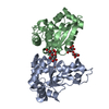
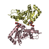
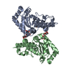
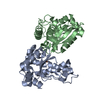
- PDB-3lj9: X-ray structure of the iron superoxide dismutase from pseudoalter... -
+ Open data

- Basic information
| Entry | Database: PDB / ID: 3lj9 | |||||||||
|---|---|---|---|---|---|---|---|---|---|---|
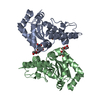
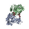
| Title | X-ray structure of the iron superoxide dismutase from pseudoalteromonas haloplanktis in complex with sodium azide | |||||||||
 Components Components | iron superoxide dismutase | |||||||||
 Keywords Keywords | OXIDOREDUCTASE / cold adaptation / superoxide dismutase / flexibility / thermal stability / psychrophilic protein / Metal-binding | |||||||||
| Function / homology |  Function and homology information Function and homology informationsuperoxide dismutase / superoxide dismutase activity / transition metal ion binding / cytoplasm Similarity search - Function | |||||||||
| Biological species |  Pseudoalteromonas haloplanktis (bacteria) Pseudoalteromonas haloplanktis (bacteria) | |||||||||
| Method |  X-RAY DIFFRACTION / MOLECULAR REPLACEMENT / Resolution: 2.1 Å X-RAY DIFFRACTION / MOLECULAR REPLACEMENT / Resolution: 2.1 Å | |||||||||
 Authors Authors | Merlino, A. / Russo Krauss, I. / Rossi, B. / Conte, M. / Vergara, A. / Sica, F. | |||||||||
 Citation Citation | Journal: J.Struct.Biol. / Year: 2010 Title: Structure and flexibility in cold-adapted iron superoxide dismutases: the case of the enzyme isolated from Pseudoalteromonas haloplanktis. Authors: Merlino, A. / Russo Krauss, I. / Castellano, I. / De Vendittis, E. / Rossi, B. / Conte, M. / Vergara, A. / Sica, F. #1: Journal: Protein and Peptide letters / Year: 2008 Title: CRYSTALLIZATION AND PRELIMINARY X-RAY DIFFRACTION STUDIES of a psychrophilic iron superoxide dismutase from Pseudoalteromonas haloplanktis Authors: Merlino, A. / Russo Krauss, I. / Castellano, I. / De Vendittis, E. / Vergara, A. / Sica, F. | |||||||||
| History |
|
- Structure visualization
| Structure viewer | Molecule: MolmilJmol/JSmol |
|---|
- Downloads & links
-Download
| PDBx/mmCIF format | 3lj9.cif.gz | 92.5 KB | Display | PDBx/mmCIF format |
|---|---|---|---|---|
| PDB format | pdb3lj9.ent.gz | 69.5 KB | Display | PDB format |
| PDBx/mmJSON format | 3lj9.json.gz | Tree view | PDBx/mmJSON format | |
| Others |  Other downloads Other downloads |
-Validation report
| Arichive directory | https://data.pdbj.org/pub/pdb/validation_reports/lj/3lj9ftp://data.pdbj.org/pub/pdb/validation_reports/lj/3lj9 | HTTPS FTP |
|---|
-Related structure data
| Related structure data |  3lioSC  3ljfC S: Starting model for refinement C: citing same article ( |
|---|---|
| Similar structure data |


-Links
 PDBj
PDBj

- Assembly
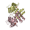
| Deposited unit | 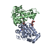
| ||||||||
|---|---|---|---|---|---|---|---|---|---|
| 1 |
| ||||||||
| Unit cell |
|
-Components
| #1: Protein | Mass: 21269.508 Da / Num. of mol.: 2 Source method: isolated from a genetically manipulated source Source: (gene. exp.) Pseudoalteromonas haloplanktis (bacteria)Strain: TAC 125 / Gene: PSHAa1215, sodB / Production host: #2: Polysaccharide | 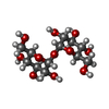  Source method: isolated from a genetically manipulated source Details: oligosaccharide with reducing-end-to-reducing-end glycosidic bond References: trehalose #3: Chemical |   Mass: 42.020 Da / Num. of mol.: 2 / Source method: obtained synthetically / Formula: N3 Mass: 42.020 Da / Num. of mol.: 2 / Source method: obtained synthetically / Formula: N3#4: Chemical |   Mass: 55.845 Da / Num. of mol.: 2 / Source method: obtained synthetically / Formula: Fe Mass: 55.845 Da / Num. of mol.: 2 / Source method: obtained synthetically / Formula: Fe#5: Water | ChemComp-HOH / |  Mass: 18.015 Da / Num. of mol.: 169 / Source method: isolated from a natural source / Formula: H2O Mass: 18.015 Da / Num. of mol.: 169 / Source method: isolated from a natural source / Formula: H2O |
|---|
-Experimental details
-Experiment
| Experiment | Method: X-RAY DIFFRACTION / Number of used crystals: 1 |
|---|
- Sample preparation
| Crystal | Density Matthews: 2.7 Å3/Da / Density % sol: 54.38 % |
|---|---|
| Crystal grow | Temperature: 293 K / Method: vapor diffusion, hanging drop / pH: 7.5 Details: 1.8M ammonium sulphate, 1.0M sodium chloride, 0.1M Hepes pH 7.5. The azide complex was prepared by soaking overnight in 2.5M ammonium sulphate, 0.1M TRIS/HCl pH 7.0., VAPOR DIFFUSION, ...Details: 1.8M ammonium sulphate, 1.0M sodium chloride, 0.1M Hepes pH 7.5. The azide complex was prepared by soaking overnight in 2.5M ammonium sulphate, 0.1M TRIS/HCl pH 7.0., VAPOR DIFFUSION, HANGING DROP, temperature 293K |
-Data collection
| Diffraction | Mean temperature: 100 K |
|---|---|
| Diffraction source | Source: ROTATING ANODE / Type: RIGAKU MICROMAX-007 HF / Wavelength: 1.5418 Å |
| Detector | Type: RIGAKU SATURN 944 / Detector: CCD / Date: Nov 27, 2007 / Details: mirrors |
| Radiation | Monochromator: GRAPHITE / Protocol: SINGLE WAVELENGTH / Monochromatic (M) / Laue (L): M / Scattering type: x-ray |
| Radiation wavelength | Wavelength: 1.5418 Å / Relative weight: 1 |
| Reflection | Resolution: 2.07→33.44 Å / Num. all: 25267 / Num. obs: 24002 / % possible obs: 97.8 % / Observed criterion σ(F): 0 / Observed criterion σ(I): 0 / Redundancy: 3.6 % / Rmerge(I) obs: 0.117 |
- Processing
| Software |
| ||||||||||||||||||||
|---|---|---|---|---|---|---|---|---|---|---|---|---|---|---|---|---|---|---|---|---|---|
| Refinement | Method to determine structure: MOLECULAR REPLACEMENT Starting model: 3LIO Resolution: 2.1→33.44 Å / Isotropic thermal model: Isotropic / Cross valid method: THROUGHOUT / σ(F): 2 / Stereochemistry target values: Engh & Huber
| ||||||||||||||||||||
| Refinement step | Cycle: LAST / Resolution: 2.1→33.44 Å
|
