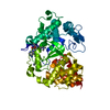
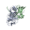
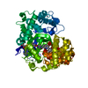
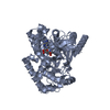
ANALYTICAL SIZE EXCLUSION CHROMATOGRAPHY SUGGESTS A DIMER AS A SIGNIFICANT OLIGOMERIZATION STATE IN SOLUTION. CRYSTAL PACKING SUPPORTS THE ASSIGNMENT OF A DIMER AS SIGNIFICANT OLIGOMERIZATION STATES.
-
Components
#1: Protein
Lipoprotein
Mass: 13551.774 Da / Num. of mol.: 2 / Fragment: sequence database residues 26-150 Source method: isolated from a genetically manipulated source Source: (gene. exp.) Shewanella oneidensis (bacteria) / Gene: SO_3163 / Plasmid: SpeedETS / Production host: Escherichia Coli (E. coli) / Strain (production host): HK100 / References: UniProt: Q8ECH9
Mass: 18.015 Da / Num. of mol.: 228 / Source method: isolated from a natural source / Formula: H2O
Has protein modification
Y
Sequence details
THE CONSTRUCT WAS EXPRESSED WITH A PURIFICATION TAG MGSDKIHHHHHHENLYFQG. THE TAG WAS REMOVED WITH ...THE CONSTRUCT WAS EXPRESSED WITH A PURIFICATION TAG MGSDKIHHHHHHENLYFQG. THE TAG WAS REMOVED WITH TEV PROTEASE LEAVING ONLY A GLYCINE (0) FOLLOWED BY THE TARGET SEQUENCE. THE CLONED CONSTRUCT CONTAINS RESIDUES 26-150 OF THE FULL LENGTH PROTEIN.
-
Experimental details
-
Experiment
Experiment
Method: X-RAY DIFFRACTION / Number of used crystals: 1
-
Sample preparation
Crystal
Density Matthews: 2.02 Å3/Da / Density % sol: 38.96 % Description: DATA WERE SCALED USING XSCALE WITH FRIEDEL PAIRS KEPT AS SEPARATE WHEN COMPUTING R-SYM, COMPLETENESS AND
Crystal grow
Temperature: 293 K / Method: vapor diffusion, sitting drop / pH: 5 Details: 1.6000M ammonium sulfate, 0.1M citric acid pH 5.0, 0.001 M copper (II) chloride, NANODROP, VAPOR DIFFUSION, SITTING DROP, temperature 293K
Monochromator: Single crystal Si(111) bent monochromator (horizontal focusing) Protocol: MAD / Monochromatic (M) / Laue (L): M / Scattering type: x-ray
Radiation wavelength
ID
Wavelength (Å)
Relative weight
1
0.91837
1
2
0.97932
1
3
0.97882
1
Reflection
Resolution: 1.42→28.421 Å / Num. obs: 43813 / % possible obs: 98.7 % / Observed criterion σ(I): -3 / Biso Wilson estimate: 16.807 Å2 / Rmerge(I) obs: 0.035 / Net I/σ(I): 18.52
Reflection shell
Resolution (Å)
Rmerge(I) obs
Mean I/σ(I) obs
Num. measured obs
Num. unique obs
Diffraction-ID
% possible all
1.42-1.47
0.492
1.7
16359
7469
1
94.3
1.47-1.53
0.347
2.8
23881
8004
1
98.3
1.53-1.6
0.253
4.4
29864
7951
1
99.3
1.6-1.68
0.178
6.2
28647
7606
1
99.3
1.68-1.79
0.122
9
31426
8308
1
99.3
1.79-1.93
0.072
14.6
30540
8066
1
99.5
1.93-2.12
0.046
22.4
29559
7724
1
99.6
2.12-2.43
0.035
30.2
30838
8033
1
99.5
2.43-3.06
0.027
39.2
30622
7951
1
99.6
3.06-28.421
0.019
53.5
30908
7996
1
98.8
-
Phasing
Phasing
Method: MAD
-
Processing
Software
Name
Version
Classification
NB
REFMAC
5.5.0053
refinement
PHENIX
refinement
SOLVE
phasing
MolProbity
3beta29
modelbuilding
XSCALE
datascaling
PDB_EXTRACT
3.006
dataextraction
XDS
datareduction
Refinement
Method to determine structure: MAD / Resolution: 1.42→28.421 Å / Cor.coef. Fo:Fc: 0.966 / Cor.coef. Fo:Fc free: 0.967 / Occupancy max: 1 / Occupancy min: 0.07 / SU B: 2.231 / SU ML: 0.039 / TLS residual ADP flag: LIKELY RESIDUAL / Cross valid method: THROUGHOUT / σ(F): 0 / ESU R: 0.063 / ESU R Free: 0.059 Stereochemistry target values: MAXIMUM LIKELIHOOD WITH PHASES Details: 1.HYDROGENS HAVE BEEN ADDED IN THE RIDING POSITIONS. 2. ATOM RECORD CONTAINS RESIDUAL B FACTORS ONLY. 3. A MET-INHIBITION PROTOCOL WAS USED FOR SELENOMETHIONINE INCORPORATION DURING PROTEIN ...Details: 1.HYDROGENS HAVE BEEN ADDED IN THE RIDING POSITIONS. 2. ATOM RECORD CONTAINS RESIDUAL B FACTORS ONLY. 3. A MET-INHIBITION PROTOCOL WAS USED FOR SELENOMETHIONINE INCORPORATION DURING PROTEIN EXPRESSION. THE OCCUPANCY OF THE SE ATOMS IN THE MSE RESIDUES WAS REDUCED TO 0.75 TO ACCOUNT FOR THE REDUCED SCATTERING POWER DUE TO PARTIAL S-MET INCORPORATION. 4.A SULFATE ION(SO4) FROM CRYSTALLIZATION AND GLYCEROL MOLECULES(GOL) FROM CRYOPROTECTANT ARE MODELED IN THE STRUCTURE.
Rfactor
Num. reflection
% reflection
Selection details
Rfree
0.181
2191
5 %
RANDOM
Rwork
0.172
-
-
-
obs
0.173
43647
99.29 %
-
Solvent computation
Ion probe radii: 0.8 Å / Shrinkage radii: 0.8 Å / VDW probe radii: 1.2 Å / Solvent model: MASK
In the structure databanks used in Yorodumi, some data are registered as the other names, "COVID-19 virus" and "2019-nCoV". Here are the details of the virus and the list of structure data.
Jan 31, 2019. EMDB accession codes are about to change! (news from PDBe EMDB page)
EMDB accession codes are about to change! (news from PDBe EMDB page)
The allocation of 4 digits for EMDB accession codes will soon come to an end. Whilst these codes will remain in use, new EMDB accession codes will include an additional digit and will expand incrementally as the available range of codes is exhausted. The current 4-digit format prefixed with “EMD-” (i.e. EMD-XXXX) will advance to a 5-digit format (i.e. EMD-XXXXX), and so on. It is currently estimated that the 4-digit codes will be depleted around Spring 2019, at which point the 5-digit format will come into force.
The EM Navigator/Yorodumi systems omit the EMD- prefix.
Related info.:Q: What is EMD? / ID/Accession-code notation in Yorodumi/EM Navigator
Yorodumi is a browser for structure data from EMDB, PDB, SASBDB, etc.
This page is also the successor to EM Navigator detail page, and also detail information page/front-end page for Omokage search.
The word "yorodu" (or yorozu) is an old Japanese word meaning "ten thousand". "mi" (miru) is to see.
Related info.:EMDB / PDB / SASBDB / Comparison of 3 databanks / Yorodumi Search / Aug 31, 2016. New EM Navigator & Yorodumi / Yorodumi Papers / Jmol/JSmol / Function and homology information / Changes in new EM Navigator and Yorodumi
 Movie
Movie Controller

 Yorodumi
 Open data
 Basic information
 Components
 Keywords
 Function and homology information
 Shewanella oneidensis (bacteria)
 X-RAY DIFFRACTION /
  Authors
 Citation
 Structure visualization
 Downloads & links
 Other downloads
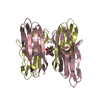
 PDBj
PDBj


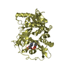
 Assembly


 Mass: 96.063 Da / Num. of mol.: 1 / Source method: obtained synthetically / Formula: SO4
 Mass: 92.094 Da / Num. of mol.: 2 / Source method: obtained synthetically / Formula: C3H8O3
 Mass: 18.015 Da / Num. of mol.: 228 / Source method: isolated from a natural source / Formula: H2O
 Sample preparation
 / Beamline: BL11-1 / Wavelength: 0.91837,0.97932,0.97882
 Processing