 Movie
Movie Controller

[English] 日本語
 Yorodumi
- PDB-3l9p: Crystal Structure of the Anaplastic Lymphoma Kinase Catalytic Domain -
+ Open data

- Basic information
| Entry | Database: PDB / ID: 3l9p | ||||||
|---|---|---|---|---|---|---|---|
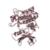
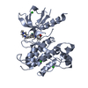
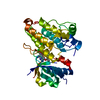
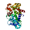
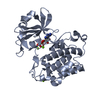
| Title | Crystal Structure of the Anaplastic Lymphoma Kinase Catalytic Domain | ||||||
 Components Components | Anaplastic lymphoma kinase | ||||||
 Keywords Keywords | TRANSFERASE / kinase domain / ATP-binding / Glycoprotein / Kinase / Membrane / Nucleotide-binding / Phosphoprotein / Proto-oncogene / Receptor / Transmembrane / Tyrosine-protein kinase | ||||||
| Function / homology |  Function and homology information Function and homology informationASP-3026-resistant ALK mutants / NVP-TAE684-resistant ALK mutants / alectinib-resistant ALK mutants / brigatinib-resistant ALK mutants / ceritinib-resistant ALK mutants / crizotinib-resistant ALK mutants / lorlatinib-resistant ALK mutants / MDK and PTN in ALK signaling / receptor signaling protein tyrosine kinase activator activity / regulation of dopamine receptor signaling pathway ...ASP-3026-resistant ALK mutants / NVP-TAE684-resistant ALK mutants / alectinib-resistant ALK mutants / brigatinib-resistant ALK mutants / ceritinib-resistant ALK mutants / crizotinib-resistant ALK mutants / lorlatinib-resistant ALK mutants / MDK and PTN in ALK signaling / receptor signaling protein tyrosine kinase activator activity / regulation of dopamine receptor signaling pathway / response to environmental enrichment / ALK mutants bind TKIs / peptidyl-tyrosine autophosphorylation / phosphorylation / positive regulation of dendrite development / regulation of neuron differentiation / Signaling by ALK / adult behavior / response to stress / negative regulation of lipid catabolic process / neuron development / energy homeostasis / swimming behavior / transmembrane receptor protein tyrosine kinase activity / cell surface receptor protein tyrosine kinase signaling pathway / : / hippocampus development / receptor protein-tyrosine kinase / Signaling by ALK fusions and activated point mutants / protein autophosphorylation / regulation of cell population proliferation / heparin binding / protein tyrosine kinase activity / regulation of apoptotic process / signaling receptor complex / signal transduction / protein-containing complex / extracellular exosome / ATP binding / identical protein binding / plasma membrane Similarity search - Function | ||||||
| Biological species |  Homo sapiens (human) Homo sapiens (human) | ||||||
| Method |  X-RAY DIFFRACTION / SYNCHROTRON / MOLECULAR REPLACEMENT / Resolution: 1.8 Å X-RAY DIFFRACTION / SYNCHROTRON / MOLECULAR REPLACEMENT / Resolution: 1.8 Å | ||||||
 Authors Authors | Lee, C. | ||||||
 Citation Citation | Journal: Biochem.J. / Year: 2010 Title: Crystal structure of the ALK (anaplastic lymphoma kinase) catalytic domain. Authors: Lee, C.C. / Jia, Y. / Li, N. / Sun, X. / Ng, K. / Ambing, E. / Gao, M.Y. / Hua, S. / Chen, C. / Kim, S. / Michellys, P.Y. / Lesley, S.A. / Harris, J.L. / Spraggon, G. | ||||||
| History |
|
- Structure visualization
| Structure viewer | Molecule: MolmilJmol/JSmol |
|---|
- Downloads & links
-Download
| PDBx/mmCIF format | 3l9p.cif.gz | 79.4 KB | Display | PDBx/mmCIF format |
|---|---|---|---|---|
| PDB format | pdb3l9p.ent.gz | 57.3 KB | Display | PDB format |
| PDBx/mmJSON format | 3l9p.json.gz | Tree view | PDBx/mmJSON format | |
| Others |  Other downloads Other downloads |
-Validation report
| Arichive directory | https://data.pdbj.org/pub/pdb/validation_reports/l9/3l9pftp://data.pdbj.org/pub/pdb/validation_reports/l9/3l9p | HTTPS FTP |
|---|
-Related structure data
| Related structure data | 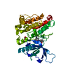 3lcsC  3lctC  1p4oS S: Starting model for refinement C: citing same article ( |
|---|---|
| Similar structure data |


-Links
 PDBj
PDBj


- Assembly
| Deposited unit | 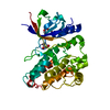
| ||||||||
|---|---|---|---|---|---|---|---|---|---|
| 1 |
| ||||||||
| Unit cell |
|
-Components
| #1: Protein | Mass: 41615.500 Da / Num. of mol.: 1 / Fragment: CATALYTIC DOMAIN residues 1072-1410 / Mutation: S1281G Source method: isolated from a genetically manipulated source Source: (gene. exp.) Homo sapiens (human) / Gene: ALK / Production host:   Spodoptera frugiperda (fall armyworm) Spodoptera frugiperda (fall armyworm)References: UniProt: Q9UM73, receptor protein-tyrosine kinase | ||
|---|---|---|---|
| #2: Chemical | ChemComp-GOL /   Mass: 92.094 Da / Num. of mol.: 4 / Source method: obtained synthetically / Formula: C3H8O3 Mass: 92.094 Da / Num. of mol.: 4 / Source method: obtained synthetically / Formula: C3H8O3#3: Water | ChemComp-HOH / |  Mass: 18.015 Da / Num. of mol.: 157 / Source method: isolated from a natural source / Formula: H2O Mass: 18.015 Da / Num. of mol.: 157 / Source method: isolated from a natural source / Formula: H2O |
-Experimental details
-Experiment
| Experiment | Method: X-RAY DIFFRACTION / Number of used crystals: 1 |
|---|
- Sample preparation
| Crystal | Density Matthews: 2.01 Å3/Da / Density % sol: 33.29 % |
|---|---|
| Crystal grow | Temperature: 277.15 K / Method: vapor diffusion, sitting drop / pH: 7.5 Details: 20% PEG 8000, 0.1M HEPEs pH 7.5, VAPOR DIFFUSION, SITTING DROP, temperature 277.15K |
-Data collection
| Diffraction | Mean temperature: 100 K |
|---|---|
| Diffraction source | Source: SYNCHROTRON / Site: ALS  / Beamline: 5.0.3 / Wavelength: 1 Å / Beamline: 5.0.3 / Wavelength: 1 Å |
| Detector | Type: ADSC QUANTUM 4 / Detector: CCD / Date: Feb 26, 2007 |
| Radiation | Protocol: SINGLE WAVELENGTH / Monochromatic (M) / Laue (L): M / Scattering type: x-ray |
| Radiation wavelength | Wavelength: 1 Å / Relative weight: 1 |
| Reflection | Resolution: 1.8→50 Å / Num. obs: 27817 / % possible obs: 95.5 % / Redundancy: 3.3 % / Rmerge(I) obs: 0.065 / Rsym value: 0.065 / Net I/σ(I): 22.17 |
| Reflection shell | Resolution: 1.8→1.83 Å / Redundancy: 1.8 % / Rmerge(I) obs: 0.065 / Rsym value: 0.065 / % possible all: 62.8 |
- Processing
| Software |
| |||||||||||||||||||||||||||||||||||||||||||||||||||||||||||||||||
|---|---|---|---|---|---|---|---|---|---|---|---|---|---|---|---|---|---|---|---|---|---|---|---|---|---|---|---|---|---|---|---|---|---|---|---|---|---|---|---|---|---|---|---|---|---|---|---|---|---|---|---|---|---|---|---|---|---|---|---|---|---|---|---|---|---|---|
| Refinement | Method to determine structure: MOLECULAR REPLACEMENT Starting model: PDB ENTRY 1P4O Resolution: 1.8→51.78 Å / Cor.coef. Fo:Fc: 0.953 / Cor.coef. Fo:Fc free: 0.934 / SU B: 2.532 / SU ML: 0.081 / Cross valid method: THROUGHOUT / ESU R: 0.142 / ESU R Free: 0.132 / Stereochemistry target values: MAXIMUM LIKELIHOOD
| |||||||||||||||||||||||||||||||||||||||||||||||||||||||||||||||||
| Solvent computation | Ion probe radii: 0.8 Å / Shrinkage radii: 0.8 Å / VDW probe radii: 1.4 Å / Solvent model: MASK | |||||||||||||||||||||||||||||||||||||||||||||||||||||||||||||||||
| Displacement parameters | Biso mean: 24.853 Å2
| |||||||||||||||||||||||||||||||||||||||||||||||||||||||||||||||||
| Refinement step | Cycle: LAST / Resolution: 1.8→51.78 Å
| |||||||||||||||||||||||||||||||||||||||||||||||||||||||||||||||||
| Refine LS restraints |
| |||||||||||||||||||||||||||||||||||||||||||||||||||||||||||||||||
| LS refinement shell | Resolution: 1.803→1.85 Å / Total num. of bins used: 20
|
