 Movie
Movie Controller

[English] 日本語
 Yorodumi
- PDB-3l9d: The Crystal Structure of smu.1046c from Streptococcus mutans UA159 -
+ Open data

- Basic information
| Entry | Database: PDB / ID: 3l9d | ||||||
|---|---|---|---|---|---|---|---|
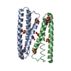
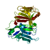
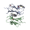
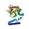
| Title | The Crystal Structure of smu.1046c from Streptococcus mutans UA159 | ||||||
 Components Components | Putative GTP pyrophosphokinase | ||||||
 Keywords Keywords | TRANSFERASE / GTP pyrophosphokinase / Kinase | ||||||
| Function / homology |  Function and homology information Function and homology informationguanosine tetraphosphate biosynthetic process / kinase activity / ATP binding Similarity search - Function | ||||||
| Biological species |  Streptococcus mutans (bacteria) Streptococcus mutans (bacteria) | ||||||
| Method |  X-RAY DIFFRACTION / MOLECULAR REPLACEMENT / Resolution: 2.484 Å X-RAY DIFFRACTION / MOLECULAR REPLACEMENT / Resolution: 2.484 Å | ||||||
 Authors Authors | Su, X.-D. / Huang, Y.H. / Liu, X. | ||||||
 Citation Citation | Journal: TO BE PUBLISHED Title: The Crystal Structure of smu.1046c from Streptococcus mutans UA159 Authors: Su, X.-D. / Huang, Y.H. / Liu, X. | ||||||
| History |
|
- Structure visualization
| Structure viewer | Molecule: MolmilJmol/JSmol |
|---|
- Downloads & links
-Download
| PDBx/mmCIF format | 3l9d.cif.gz | 95.6 KB | Display | PDBx/mmCIF format |
|---|---|---|---|---|
| PDB format | pdb3l9d.ent.gz | 72.1 KB | Display | PDB format |
| PDBx/mmJSON format | 3l9d.json.gz | Tree view | PDBx/mmJSON format | |
| Others |  Other downloads Other downloads |
-Validation report
| Arichive directory | https://data.pdbj.org/pub/pdb/validation_reports/l9/3l9dftp://data.pdbj.org/pub/pdb/validation_reports/l9/3l9d | HTTPS FTP |
|---|
-Related structure data
| Related structure data |  2be3S S: Starting model for refinement |
|---|---|
| Similar structure data |


-Links
 PDBj
PDBj- Assembly
| Deposited unit | 
| ||||||||
|---|---|---|---|---|---|---|---|---|---|
| 1 | 
| ||||||||
| 2 | 
| ||||||||
| Unit cell |
| ||||||||
| Components on special symmetry positions |
|
-Components
| #1: Protein | Mass: 29876.189 Da / Num. of mol.: 2 Source method: isolated from a genetically manipulated source Source: (gene. exp.) Streptococcus mutans (bacteria) / Strain: UA159 / Gene: smu.1046c / Plasmid: pET28a / Production host: #2: Water | ChemComp-HOH / |  Mass: 18.015 Da / Num. of mol.: 128 / Source method: isolated from a natural source / Formula: H2O Mass: 18.015 Da / Num. of mol.: 128 / Source method: isolated from a natural source / Formula: H2O |
|---|
-Experimental details
-Experiment
| Experiment | Method: X-RAY DIFFRACTION / Number of used crystals: 1 |
|---|
- Sample preparation
| Crystal | Density Matthews: 2.15 Å3/Da / Density % sol: 42.73 % |
|---|---|
| Crystal grow | Temperature: 289 K / Method: vapor diffusion, sitting drop / pH: 7.5 Details: 8% PEG 1000, 0.1mM KCl, 0.1mM HEPES Na pH 7.0, 0.02mM MgCl2 , VAPOR DIFFUSION, SITTING DROP, temperature 289K |
-Data collection
| Diffraction | Mean temperature: 100 K |
|---|---|
| Diffraction source | Source: ROTATING ANODE / Type: BRUKER AXS MICROSTAR-H / Wavelength: 1.54 Å |
| Detector | Type: BRUKER SMART 6000 / Detector: CCD / Date: Apr 20, 2009 |
| Radiation | Protocol: SINGLE WAVELENGTH / Monochromatic (M) / Laue (L): M / Scattering type: x-ray |
| Radiation wavelength | Wavelength: 1.54 Å / Relative weight: 1 |
| Reflection | Resolution: 2.484→62.06 Å / Num. obs: 17850 / Observed criterion σ(I): 1 / Biso Wilson estimate: 31.55 Å2 |
| Reflection shell | Highest resolution: 2.484 Å |
- Processing
| Software |
| ||||||||||||||||||||||||||||||||||||||||||||||||||||||||||||||||||||||||||||||||||||
|---|---|---|---|---|---|---|---|---|---|---|---|---|---|---|---|---|---|---|---|---|---|---|---|---|---|---|---|---|---|---|---|---|---|---|---|---|---|---|---|---|---|---|---|---|---|---|---|---|---|---|---|---|---|---|---|---|---|---|---|---|---|---|---|---|---|---|---|---|---|---|---|---|---|---|---|---|---|---|---|---|---|---|---|---|---|
| Refinement | Method to determine structure: MOLECULAR REPLACEMENT Starting model: PDB ENTRY 2BE3 Resolution: 2.484→38.81 Å / Occupancy max: 1 / Occupancy min: 0.7 / FOM work R set: 0.7851 / SU ML: 0.33 / Cross valid method: THROUGHOUT / σ(F): 0.06 / Phase error: 28.32 / Stereochemistry target values: ML
| ||||||||||||||||||||||||||||||||||||||||||||||||||||||||||||||||||||||||||||||||||||
| Solvent computation | Shrinkage radii: 0.9 Å / VDW probe radii: 1.11 Å / Solvent model: FLAT BULK SOLVENT MODEL / Bsol: 33.538 Å2 / ksol: 0.334 e/Å3 | ||||||||||||||||||||||||||||||||||||||||||||||||||||||||||||||||||||||||||||||||||||
| Displacement parameters | Biso max: 102.94 Å2 / Biso mean: 37.3679 Å2 / Biso min: 11.83 Å2
| ||||||||||||||||||||||||||||||||||||||||||||||||||||||||||||||||||||||||||||||||||||
| Refinement step | Cycle: LAST / Resolution: 2.484→38.81 Å
| ||||||||||||||||||||||||||||||||||||||||||||||||||||||||||||||||||||||||||||||||||||
| Refine LS restraints |
| ||||||||||||||||||||||||||||||||||||||||||||||||||||||||||||||||||||||||||||||||||||
| LS refinement shell | Refine-ID: X-RAY DIFFRACTION / Total num. of bins used: 11
|
