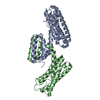
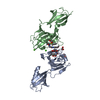
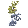
Programmedcelldeathprotein10 / TF-1 cell apoptosis-related protein 15 / Cerebral cavernous malformations 3 protein
Mass: 23376.994 Da / Num. of mol.: 1 Source method: isolated from a genetically manipulated source Source: (gene. exp.) Homo sapiens (human) / Gene: PDCD10, CCM3, TFAR15 / Plasmid: modified pET-32 / Production host: Escherichia coli (E. coli) / Strain (production host): Rosetta (DE3) / References: UniProt: Q9BUL8
Sequence details
RESIDUES -3, -2 AND -1 ARE BUILT INTO POOR DENSITY, RESIDUE ASSIGNMENT IS UNCLEAR. THE COMPLETE ...RESIDUES -3, -2 AND -1 ARE BUILT INTO POOR DENSITY, RESIDUE ASSIGNMENT IS UNCLEAR. THE COMPLETE CRYSTALLIZED SEQUENCE IS: GHMRMTMEEMKNEAETTSMVSMPLYAVMYPVFNELERVNLSAAQTLRAAFIKAEKENPGLTQDIIMKILEKKSVEVNFTESL LRMAADDVEEYMIERPEPEFQDLNEKARALKQILSKIPDEINDRVRFLQTIKDIASAIKELLDTVNNVFKKYQYQNRRALEH QKKEFVKYSKSFSDTLKTYFKDGKAINVFVSANRLIHQTNLILQTFKTVA
-
Experimental details
-
Experiment
Experiment
Method: X-RAY DIFFRACTION / Number of used crystals: 1
-
Sample preparation
Crystal
Density Matthews: 3.48 Å3/Da / Density % sol: 64.69 %
Crystal grow
Temperature: 298 K / Method: vapor diffusion, hanging drop / pH: 6.5 Details: 0.16 M calcium acetate, 0.08 M sodium cacodylate pH 6.5, 20% glycerol, 13.5% PEG 8000, VAPOR DIFFUSION, HANGING DROP, temperature 298K
Resolution: 3.05→25 Å / Cor.coef. Fo:Fc: 0.942 / Cor.coef. Fo:Fc free: 0.934 / SU B: 70.715 / SU ML: 0.532 / Cross valid method: THROUGHOUT / ESU R Free: 0.472 / Stereochemistry target values: MAXIMUM LIKELIHOOD Details: HYDROGENS HAVE BEEN ADDED IN THE RIDING POSITIONS. Residues -3, -2 and -1 are built into poor density, residue assignment is unclear.
Rfactor
Num. reflection
% reflection
Selection details
Rfree
0.2926
316
4.9 %
RANDOM
Rwork
0.27827
-
-
-
obs
0.27895
6170
96.56 %
-
Solvent computation
Ion probe radii: 0.8 Å / Shrinkage radii: 0.8 Å / VDW probe radii: 1.4 Å / Solvent model: MASK
Displacement parameters
Biso mean: 146.451 Å2
Baniso -1
Baniso -2
Baniso -3
1-
5.61 Å2
0 Å2
0 Å2
2-
-
5.61 Å2
0 Å2
3-
-
-
-11.23 Å2
Refinement step
Cycle: LAST / Resolution: 3.05→25 Å
Protein
Nucleic acid
Ligand
Solvent
Total
Num. atoms
1630
0
0
0
1630
Refine LS restraints
Refine-ID
Type
Dev ideal
Dev ideal target
Number
X-RAY DIFFRACTION
r_bond_refined_d
0.006
0.022
1654
X-RAY DIFFRACTION
r_bond_other_d
X-RAY DIFFRACTION
r_angle_refined_deg
0.881
1.972
2224
X-RAY DIFFRACTION
r_angle_other_deg
X-RAY DIFFRACTION
r_dihedral_angle_1_deg
3.899
5
198
X-RAY DIFFRACTION
r_dihedral_angle_2_deg
37.883
24.937
79
X-RAY DIFFRACTION
r_dihedral_angle_3_deg
15.178
15
329
X-RAY DIFFRACTION
r_dihedral_angle_4_deg
13.058
15
10
X-RAY DIFFRACTION
r_chiral_restr
0.054
0.2
256
X-RAY DIFFRACTION
r_gen_planes_refined
0.003
0.021
1207
X-RAY DIFFRACTION
r_gen_planes_other
X-RAY DIFFRACTION
r_nbd_refined
X-RAY DIFFRACTION
r_nbd_other
X-RAY DIFFRACTION
r_nbtor_refined
X-RAY DIFFRACTION
r_nbtor_other
X-RAY DIFFRACTION
r_xyhbond_nbd_refined
X-RAY DIFFRACTION
r_xyhbond_nbd_other
X-RAY DIFFRACTION
r_metal_ion_refined
X-RAY DIFFRACTION
r_metal_ion_other
X-RAY DIFFRACTION
r_symmetry_vdw_refined
X-RAY DIFFRACTION
r_symmetry_vdw_other
X-RAY DIFFRACTION
r_symmetry_hbond_refined
X-RAY DIFFRACTION
r_symmetry_hbond_other
X-RAY DIFFRACTION
r_symmetry_metal_ion_refined
X-RAY DIFFRACTION
r_symmetry_metal_ion_other
X-RAY DIFFRACTION
r_mcbond_it
0.106
1.5
1002
X-RAY DIFFRACTION
r_mcbond_other
X-RAY DIFFRACTION
r_mcangle_it
0.199
2
1630
X-RAY DIFFRACTION
r_scbond_it
0.228
3
652
X-RAY DIFFRACTION
r_scangle_it
0.4
4.5
594
X-RAY DIFFRACTION
r_rigid_bond_restr
X-RAY DIFFRACTION
r_sphericity_free
X-RAY DIFFRACTION
r_sphericity_bonded
LS refinement shell
Resolution: 3.05→3.128 Å / Total num. of bins used: 20
Rfactor
Num. reflection
% reflection
Rfree
0.415
30
-
Rwork
0.349
420
-
obs
-
-
95.34 %
Refinement TLS params.
Method: refined / Refine-ID: X-RAY DIFFRACTION
ID
L11 (°2)
L12 (°2)
L13 (°2)
L22 (°2)
L23 (°2)
L33 (°2)
S11 (Å °)
S12 (Å °)
S13 (Å °)
S21 (Å °)
S22 (Å °)
S23 (Å °)
S31 (Å °)
S32 (Å °)
S33 (Å °)
T11 (Å2)
T12 (Å2)
T13 (Å2)
T22 (Å2)
T23 (Å2)
T33 (Å2)
Origin x (Å)
Origin y (Å)
Origin z (Å)
1
10.2247
3.1313
6.266
11.0703
0.6746
14.9114
1.1338
0.2457
-0.2971
0.2081
0.0414
-0.3427
2.2397
1.8038
-1.1753
0.3899
0.3363
-0.2013
0.4479
-0.2785
0.2035
54.2645
27.384
19.2442
2
12.8797
4.8837
-2.8661
8.3192
-4.9042
7.0042
-0.2006
-0.3794
-0.6435
-0.4162
1.0693
1.2207
0.128
-0.8815
-0.8687
0.3478
0.1576
-0.1018
0.5535
0.4663
0.5892
20.8587
19.9686
14.6578
Refinement TLS group
ID
Refine-ID
Refine TLS-ID
Auth asym-ID
Auth seq-ID
1
X-RAY DIFFRACTION
1
A
14 - 69
2
X-RAY DIFFRACTION
2
A
70 - 210
+
About Yorodumi
-
News
-
Feb 9, 2022. New format data for meta-information of EMDB entries
New format data for meta-information of EMDB entries
Version 3 of the EMDB header file is now the official format.
The previous official version 1.9 will be removed from the archive.
In the structure databanks used in Yorodumi, some data are registered as the other names, "COVID-19 virus" and "2019-nCoV". Here are the details of the virus and the list of structure data.
Jan 31, 2019. EMDB accession codes are about to change! (news from PDBe EMDB page)
EMDB accession codes are about to change! (news from PDBe EMDB page)
The allocation of 4 digits for EMDB accession codes will soon come to an end. Whilst these codes will remain in use, new EMDB accession codes will include an additional digit and will expand incrementally as the available range of codes is exhausted. The current 4-digit format prefixed with “EMD-” (i.e. EMD-XXXX) will advance to a 5-digit format (i.e. EMD-XXXXX), and so on. It is currently estimated that the 4-digit codes will be depleted around Spring 2019, at which point the 5-digit format will come into force.
The EM Navigator/Yorodumi systems omit the EMD- prefix.
Related info.:Q: What is EMD? / ID/Accession-code notation in Yorodumi/EM Navigator
Yorodumi is a browser for structure data from EMDB, PDB, SASBDB, etc.
This page is also the successor to EM Navigator detail page, and also detail information page/front-end page for Omokage search.
The word "yorodu" (or yorozu) is an old Japanese word meaning "ten thousand". "mi" (miru) is to see.
Related info.:EMDB / PDB / SASBDB / Comparison of 3 databanks / Yorodumi Search / Aug 31, 2016. New EM Navigator & Yorodumi / Yorodumi Papers / Jmol/JSmol / Function and homology information / Changes in new EM Navigator and Yorodumi
 Movie
Movie Controller

 Yorodumi
 Open data
 Basic information
 Components
 Keywords
 Function and homology information
 Homo sapiens (human)
 X-RAY DIFFRACTION /
  Authors
 Citation
 Structure visualization
 Downloads & links
 Other downloads


 PDBj
PDBj Assembly
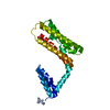

 Sample preparation
 / Beamline: X6A / Wavelength: 0.9551 Å
 Processing