 Movie
Movie Controller

[English] 日本語
 Yorodumi
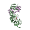
- PDB-3l6w: Structure of the collar functional unit (KLH1-H) of keyhole limpe... -
+ Open data

- Basic information
| Entry | Database: PDB / ID: 3l6w | ||||||
|---|---|---|---|---|---|---|---|
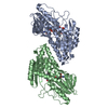
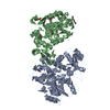
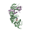
| Title | Structure of the collar functional unit (KLH1-H) of keyhole limpet hemocyanin | ||||||
 Components Components | Hemocyanin 1 | ||||||
 Keywords Keywords | OXYGEN BINDING / HEMOCYANIN / CUPREDOXIN DOMAIN / COPPER-BINDING PROTEIN / METAL-BINDING | ||||||
| Function / homology |  Function and homology information Function and homology informationoxygen carrier activity / oxidoreductase activity / extracellular region / metal ion binding Similarity search - Function | ||||||
| Biological species |  Megathura crenulata (invertebrata) Megathura crenulata (invertebrata) | ||||||
| Method |  X-RAY DIFFRACTION / SYNCHROTRON / MOLECULAR REPLACEMENT / Resolution: 4 Å X-RAY DIFFRACTION / SYNCHROTRON / MOLECULAR REPLACEMENT / Resolution: 4 Å | ||||||
 Authors Authors | Jaenicke, E. / Buechler, K. / Markl, J. / Decker, H. / Barends, T.R.M. | ||||||
 Citation Citation | Journal: Biochem.J. / Year: 2009 Title: The Cupredoxin-like Domains in Hemocyanins. Authors: Jaenicke, E. / Buchler, K. / Markl, J. / Decker, H. / Barends, T.R.M. | ||||||
| History |
|
- Structure visualization
| Structure viewer | Molecule: MolmilJmol/JSmol |
|---|
- Downloads & links
-Download
| PDBx/mmCIF format | 3l6w.cif.gz | 40.8 KB | Display | PDBx/mmCIF format |
|---|---|---|---|---|
| PDB format | pdb3l6w.ent.gz | 21.9 KB | Display | PDB format |
| PDBx/mmJSON format | 3l6w.json.gz | Tree view | PDBx/mmJSON format | |
| Others |  Other downloads Other downloads |
-Validation report
| Arichive directory | https://data.pdbj.org/pub/pdb/validation_reports/l6/3l6wftp://data.pdbj.org/pub/pdb/validation_reports/l6/3l6w | HTTPS FTP |
|---|
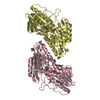
-Related structure data
| Related structure data | 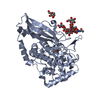 1js8S  3eu2 S: Starting model for refinement |
|---|---|
| Similar structure data |


-Links
 PDBj
PDBj- Assembly
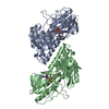
| Deposited unit | 
| ||||||||
|---|---|---|---|---|---|---|---|---|---|
| 1 |
| ||||||||
| Unit cell |
|
-Components
| #1: Protein | Mass: 56623.797 Da / Num. of mol.: 2 / Fragment: collar subunit / Source method: isolated from a natural source / Source: (natural) Megathura crenulata (invertebrata) / References: UniProt: Q53IP9, UniProt: Q10583*PLUS |
|---|
-Experimental details
-Experiment
| Experiment | Method: X-RAY DIFFRACTION / Number of used crystals: 1 |
|---|
- Sample preparation
| Crystal | Density Matthews: 5.82 Å3/Da / Density % sol: 78.86 % |
|---|---|
| Crystal grow | Temperature: 293 K / Method: vapor diffusion, sitting drop / pH: 4 Details: 9% PEG 1000, 50mM phosphate/citrate buffer, pH 4.0, VAPOR DIFFUSION, SITTING DROP, temperature 293K |
-Data collection
| Diffraction | Mean temperature: 90 K |
|---|---|
| Diffraction source | Source: SYNCHROTRON / Site: SLS  / Beamline: X06SA / Wavelength: 0.97941 / Beamline: X06SA / Wavelength: 0.97941 |
| Detector | Type: MARMOSAIC 225 mm CCD / Detector: CCD / Date: Sep 22, 2006 |
| Radiation | Monochromator: SI(111) / Protocol: SINGLE WAVELENGTH / Monochromatic (M) / Laue (L): M / Scattering type: x-ray |
| Radiation wavelength | Wavelength: 0.97941 Å / Relative weight: 1 |
| Reflection | Resolution: 4→30 Å / Num. obs: 22294 / % possible obs: 96.8 % / Observed criterion σ(I): 0 / Rmerge(I) obs: 0.18 / Net I/σ(I): 10.6 |
| Reflection shell | Resolution: 4→4.2 Å / Rmerge(I) obs: 0.847 / Mean I/σ(I) obs: 3.46 / % possible all: 96.4 |
- Processing
| Software |
| ||||||||||||||||||
|---|---|---|---|---|---|---|---|---|---|---|---|---|---|---|---|---|---|---|---|
| Refinement | Method to determine structure: MOLECULAR REPLACEMENT Starting model: PDB ENTRY 1JS8 Highest resolution: 4 Å Details: THE COORDINATES CONTAIN ONLY A CA TRACE. NO REFINEMENT WAS CARRIED OUT | ||||||||||||||||||
| Refinement step | Cycle: LAST / Highest resolution: 4 Å
|
