 Movie
Movie Controller

[English] 日本語
 Yorodumi
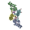
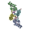
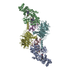
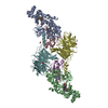
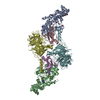
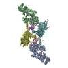
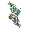
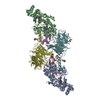
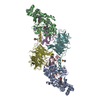
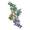
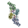
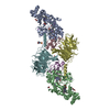
- PDB-3l4o: Crystal Structure of the MauG/pre-Methylamine Dehydrogenase Compl... -
+ Open data

- Basic information
| Entry | Database: PDB / ID: 3l4o | ||||||
|---|---|---|---|---|---|---|---|
| Title | Crystal Structure of the MauG/pre-Methylamine Dehydrogenase Complex After Treatment with Hydrogen Peroxide | ||||||
 Components Components |
| ||||||
 Keywords Keywords | OXIDOREDUCTASE/ELECTRON TRANSPORT / MauG / methylamine dehydrogenase / quinone cofactor / TTQ / His-Tyr heme / electron transport / c-heme / Iron / metal-binding / Oxidoreductase / Transport / Disulfide bond / OXIDOREDUCTASE-ELECTRON TRANSPORT complex | ||||||
| Function / homology |  Function and homology information Function and homology informationmethylamine dehydrogenase (amicyanin) / methylamine dehydrogenase (amicyanin) activity / methylamine metabolic process / aliphatic amine dehydrogenase activity / amine metabolic process / Oxidoreductases / cytochrome-c peroxidase activity / outer membrane-bounded periplasmic space / periplasmic space / electron transfer activity ...methylamine dehydrogenase (amicyanin) / methylamine dehydrogenase (amicyanin) activity / methylamine metabolic process / aliphatic amine dehydrogenase activity / amine metabolic process / Oxidoreductases / cytochrome-c peroxidase activity / outer membrane-bounded periplasmic space / periplasmic space / electron transfer activity / heme binding / metal ion binding Similarity search - Function | ||||||
| Biological species |  Paracoccus denitrificans (bacteria) Paracoccus denitrificans (bacteria) | ||||||
| Method |  X-RAY DIFFRACTION / SYNCHROTRON / Difference Fourier / molecular replacement / Resolution: 2.046 Å X-RAY DIFFRACTION / SYNCHROTRON / Difference Fourier / molecular replacement / Resolution: 2.046 Å | ||||||
 Authors Authors | Jensen, L.M.R. / Wilmot, C.M. | ||||||
 Citation Citation | Journal: Science / Year: 2010 Title: In crystallo posttranslational modification within a MauG/pre-methylamine dehydrogenase complex. Authors: Jensen, L.M. / Sanishvili, R. / Davidson, V.L. / Wilmot, C.M. | ||||||
| History |
|
- Structure visualization
| Structure viewer | Molecule: MolmilJmol/JSmol |
|---|
- Downloads & links
-Download
| PDBx/mmCIF format | 3l4o.cif.gz | 374.6 KB | Display | PDBx/mmCIF format |
|---|---|---|---|---|
| PDB format | pdb3l4o.ent.gz | 299.1 KB | Display | PDB format |
| PDBx/mmJSON format | 3l4o.json.gz | Tree view | PDBx/mmJSON format | |
| Others |  Other downloads Other downloads |
-Validation report
| Summary document | 3l4o_validation.pdf.gz | 1.9 MB | Display | wwPDB validaton report |
|---|---|---|---|---|
| Full document | 3l4o_full_validation.pdf.gz | 1.9 MB | Display | |
| Data in XML | 3l4o_validation.xml.gz | 76 KB | Display | |
| Data in CIF | 3l4o_validation.cif.gz | 109.6 KB | Display | |
| Arichive directory | https://data.pdbj.org/pub/pdb/validation_reports/l4/3l4oftp://data.pdbj.org/pub/pdb/validation_reports/l4/3l4o | HTTPS FTP |
-Related structure data
| Related structure data |  3l4mSC S: Starting model for refinement C: citing same article ( |
|---|---|
| Similar structure data |


-Links
 PDBj
PDBj


- Assembly
| Deposited unit | 
| ||||||||
|---|---|---|---|---|---|---|---|---|---|
| 1 |
| ||||||||
| Unit cell |
|
-Components
-Protein / Antibody / Methylamine dehydrogenase ... , 3 types, 6 molecules ABCEDF
| #1: Protein | Mass: 41146.629 Da / Num. of mol.: 2 Source method: isolated from a genetically manipulated source Source: (gene. exp.) Paracoccus denitrificans (bacteria) / Strain: Pd 1222 / Gene: mauG / Production host: Paracoccus denitrificans (bacteria) / References: UniProt: Q51658, Oxidoreductases#2: Antibody | Mass: 15039.577 Da / Num. of mol.: 2 Fragment: Beta chain of immature methylamine dehydrogenase (preMADH) Mutation: Hydroxylated Trp57 has been converted to the full-quinone form due to hydrogen peroxide-assisted catalysis by MauG in the crystal. Source method: isolated from a genetically manipulated source Details: Immature MADH (preMADH) was produced in the absence of the mauG gene. Trp57 is monohydroxylated at the C7 position. Source: (gene. exp.) Paracoccus denitrificans (bacteria) / Strain: Pd 1222 / Gene: mauA / Production host: Rhodobacter sphaeroides (bacteria)References: UniProt: P22619, UniProt: A1BBA0*PLUS, EC: 1.4.99.3 #3: Protein | Mass: 42449.277 Da / Num. of mol.: 2 Source method: isolated from a genetically manipulated source Details: Immature MADH (preMADH) was produced in the absence of the mauG gene. The alpha subunit has the wild-type sequence. Source: (gene. exp.) Paracoccus denitrificans (bacteria) / Strain: Pd 1222 / Gene: Pden_4730 / Production host: Rhodobacter sphaeroides (bacteria) / References: UniProt: A1BB97, EC: 1.4.99.3 |
|---|
-Non-polymers , 6 types, 1277 molecules 


| #4: Chemical |  Mass: 40.078 Da / Num. of mol.: 2 / Source method: obtained synthetically / Formula: Ca Mass: 40.078 Da / Num. of mol.: 2 / Source method: obtained synthetically / Formula: Ca#5: Chemical | ChemComp-HEC /  Mass: 618.503 Da / Num. of mol.: 4 / Source method: obtained synthetically / Formula: C34H34FeN4O4 Mass: 618.503 Da / Num. of mol.: 4 / Source method: obtained synthetically / Formula: C34H34FeN4O4#6: Chemical | ChemComp-1PE / |  Mass: 238.278 Da / Num. of mol.: 1 / Source method: obtained synthetically / Formula: C10H22O6 / Comment: precipitant*YM Mass: 238.278 Da / Num. of mol.: 1 / Source method: obtained synthetically / Formula: C10H22O6 / Comment: precipitant*YM#7: Chemical | ChemComp-PG4 / |  Mass: 194.226 Da / Num. of mol.: 1 / Source method: obtained synthetically / Formula: C8H18O5 / Comment: precipitant*YM Mass: 194.226 Da / Num. of mol.: 1 / Source method: obtained synthetically / Formula: C8H18O5 / Comment: precipitant*YM#8: Chemical | ChemComp-ACT / |  Mass: 59.044 Da / Num. of mol.: 1 / Source method: obtained synthetically / Formula: C2H3O2 Mass: 59.044 Da / Num. of mol.: 1 / Source method: obtained synthetically / Formula: C2H3O2#9: Water | ChemComp-HOH / | Mass: 18.015 Da / Num. of mol.: 1268 / Source method: isolated from a natural source / Formula: H2O |
|---|
-Experimental details
-Experiment
| Experiment | Method: X-RAY DIFFRACTION / Number of used crystals: 1 |
|---|
- Sample preparation
| Crystal | Density Matthews: 2.29 Å3/Da / Density % sol: 46.32 % |
|---|---|
| Crystal grow | Temperature: 293 K / Method: vapor diffusion, hanging drop / pH: 6.4 Details: 0.1M MES pH 6.4, 0.1M sodium acetate, 24-30 % w/v PEG 8000; followed by reaction of the crystal with hydrogen peroxide immediately prior to flash cooling., VAPOR DIFFUSION, HANGING DROP, temperature 293K |
-Data collection
| Diffraction | Mean temperature: 100 K |
|---|---|
| Diffraction source | Source: SYNCHROTRON / Site: APS  / Beamline: 23-ID-B / Wavelength: 1.0332 Å / Beamline: 23-ID-B / Wavelength: 1.0332 Å |
| Detector | Type: MARMOSAIC 300 mm CCD / Detector: CCD / Date: Aug 17, 2009 / Details: BIOMORPH MIRRORS (KIRKPATRICK-BAEZ CONFIGURATION) |
| Radiation | Monochromator: Si(111) Double crystal monochromator / Protocol: SINGLE WAVELENGTH / Scattering type: x-ray |
| Radiation wavelength | Wavelength: 1.0332 Å / Relative weight: 1 |
| Reflection | Resolution: 1.96→50 Å / Num. all: 123424 / Num. obs: 120574 / % possible obs: 97.7 % / Observed criterion σ(F): 0 / Observed criterion σ(I): -3 / Redundancy: 3.9 % / Biso Wilson estimate: 36.09 Å2 / Rmerge(I) obs: 0.075 / Rsym value: 0.075 / Χ2: 1.042 / Net I/σ(I): 17.1 |
| Reflection shell | Resolution: 1.96→1.99 Å / Redundancy: 3.2 % / Rmerge(I) obs: 0.524 / Mean I/σ(I) obs: 2.1 / Num. unique all: 5673 / Χ2: 1.096 / % possible all: 92.5 |
-Phasing
| Phasing | Method: molecular replacement |
|---|
- Processing
| Software |
| |||||||||||||||||||||||||||||||||||||||||||||||||||||||||||||||||||||||||||||||||||||||||||||||||||||||||||||||||||||||||||||||||||||||||||||||||||||||||||||||||||||||||||||||
|---|---|---|---|---|---|---|---|---|---|---|---|---|---|---|---|---|---|---|---|---|---|---|---|---|---|---|---|---|---|---|---|---|---|---|---|---|---|---|---|---|---|---|---|---|---|---|---|---|---|---|---|---|---|---|---|---|---|---|---|---|---|---|---|---|---|---|---|---|---|---|---|---|---|---|---|---|---|---|---|---|---|---|---|---|---|---|---|---|---|---|---|---|---|---|---|---|---|---|---|---|---|---|---|---|---|---|---|---|---|---|---|---|---|---|---|---|---|---|---|---|---|---|---|---|---|---|---|---|---|---|---|---|---|---|---|---|---|---|---|---|---|---|---|---|---|---|---|---|---|---|---|---|---|---|---|---|---|---|---|---|---|---|---|---|---|---|---|---|---|---|---|---|---|---|---|---|
| Refinement | Method to determine structure: Difference Fourier Starting model: 3l4m Resolution: 2.046→38.2 Å / Cor.coef. Fo:Fc: 0.973 / Cor.coef. Fo:Fc free: 0.952 / WRfactor Rfree: 0.197 / WRfactor Rwork: 0.146 / Occupancy max: 1 / Occupancy min: 0.3 / FOM work R set: 0.883 / SU B: 8.088 / SU ML: 0.105 / SU R Cruickshank DPI: 0.177 / SU Rfree: 0.156 / TLS residual ADP flag: LIKELY RESIDUAL / Cross valid method: THROUGHOUT / σ(F): 0 / ESU R: 0.177 / ESU R Free: 0.156 / Stereochemistry target values: MAXIMUM LIKELIHOOD Details: HYDROGENS HAVE BEEN ADDED IN THE RIDING POSITIONS U VALUES: RESIDUAL ONLY
| |||||||||||||||||||||||||||||||||||||||||||||||||||||||||||||||||||||||||||||||||||||||||||||||||||||||||||||||||||||||||||||||||||||||||||||||||||||||||||||||||||||||||||||||
| Solvent computation | Ion probe radii: 0.8 Å / Shrinkage radii: 0.8 Å / VDW probe radii: 1.4 Å / Solvent model: BABINET MODEL WITH MASK | |||||||||||||||||||||||||||||||||||||||||||||||||||||||||||||||||||||||||||||||||||||||||||||||||||||||||||||||||||||||||||||||||||||||||||||||||||||||||||||||||||||||||||||||
| Displacement parameters | Biso max: 69.61 Å2 / Biso mean: 21.828 Å2 / Biso min: 7.01 Å2
| |||||||||||||||||||||||||||||||||||||||||||||||||||||||||||||||||||||||||||||||||||||||||||||||||||||||||||||||||||||||||||||||||||||||||||||||||||||||||||||||||||||||||||||||
| Refinement step | Cycle: LAST / Resolution: 2.046→38.2 Å
| |||||||||||||||||||||||||||||||||||||||||||||||||||||||||||||||||||||||||||||||||||||||||||||||||||||||||||||||||||||||||||||||||||||||||||||||||||||||||||||||||||||||||||||||
| Refine LS restraints |
| |||||||||||||||||||||||||||||||||||||||||||||||||||||||||||||||||||||||||||||||||||||||||||||||||||||||||||||||||||||||||||||||||||||||||||||||||||||||||||||||||||||||||||||||
| LS refinement shell | Resolution: 2.046→2.099 Å / Total num. of bins used: 20
| |||||||||||||||||||||||||||||||||||||||||||||||||||||||||||||||||||||||||||||||||||||||||||||||||||||||||||||||||||||||||||||||||||||||||||||||||||||||||||||||||||||||||||||||
| Refinement TLS params. | Method: refined / Refine-ID: X-RAY DIFFRACTION
| |||||||||||||||||||||||||||||||||||||||||||||||||||||||||||||||||||||||||||||||||||||||||||||||||||||||||||||||||||||||||||||||||||||||||||||||||||||||||||||||||||||||||||||||
| Refinement TLS group |
|
