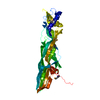
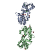
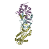
Entry Database : PDB / ID : 3l4fTitle Crystal Structure of betaPIX Coiled-Coil Domain and Shank PDZ Complex Rho guanine nucleotide exchange factor 7 SH3 and multiple ankyrin repeat domains protein 1 Keywords / / / / / / / / / / / / Function / homology Function Domain/homology Component
/ / / / / / / / / / / / / / / / / / / / / / / / / / / / / / / / / / / / / / / / / / / / / / / / / / / / / / / / / / / / / / / / / / / / / / / / / / / / / / / / / / / / / / / / / / / / / / / / / / / / / / / / / / / / / / / / / / / / / / / / / / / / / / / / / / / / / / / / / / / / / / / / / Biological species Rattus norvegicus (Norway rat)Method / / / Resolution : 2.8 Å Authors Im, Y.J. / Kang, G.B. / Lee, J.H. / Song, H.E. / Park, K.R. / Kim, E. / Song, W.K. / Park, D. / Eom, S.H. Journal : J.Mol.Biol. / Year : 2010Title : Structural basis for asymmetric association of the betaPIX coiled coil and shank PDZAuthors : Im, Y.J. / Kang, G.B. / Lee, J.H. / Park, K.R. / Song, H.E. / Kim, E. / Song, W.K. / Park, D. / Eom, S.H. History Deposition Dec 19, 2009 Deposition site / Processing site Revision 1.0 Feb 16, 2010 Provider / Type Revision 1.1 Jul 13, 2011 Group Revision 1.2 Feb 26, 2014 Group Revision 1.3 Dec 25, 2019 Group / Database references / Category / struct_ref_seq_difItem / _struct_ref_seq_dif.detailsRevision 1.4 Nov 1, 2023 Group / Database references / Refinement descriptionCategory chem_comp_atom / chem_comp_bond ... chem_comp_atom / chem_comp_bond / database_2 / pdbx_initial_refinement_model Item / _database_2.pdbx_database_accession
Show all Show less
 Movie
Movie Controller

 Yorodumi
 Open data
 Basic information
 Components
 Keywords
 Function and homology information
 X-RAY DIFFRACTION /
  Authors
 Citation
 Structure visualization
 Downloads & links
 Other downloads
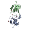


 PDBj
PDBj

 Assembly

 Mass: 18.015 Da / Num. of mol.: 20 / Source method: isolated from a natural source / Formula: H2O
 Sample preparation

 Processing