 Movie
Movie Controller

[English] 日本語
 Yorodumi
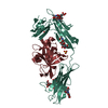
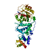
- PDB-3ktw: Crystal structure of the SRP19/S-domain SRP RNA complex of Sulfol... -
+ Open data

- Basic information
| Entry | Database: PDB / ID: 3ktw | ||||||
|---|---|---|---|---|---|---|---|
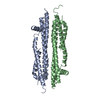
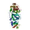
| Title | Crystal structure of the SRP19/S-domain SRP RNA complex of Sulfolobus solfataricus | ||||||
 Components Components |
| ||||||
 Keywords Keywords | RNA/RNA binding protein / ribonucleoprotein complex / RNA-RNA tertiary interactions / asymmetric loop / 7S RNA / signal recognition particle / structural RNA / RNA-binding / RNA-RNA binding protein complex | ||||||
| Function / homology |  Function and homology information Function and homology informationsignal recognition particle / 7S RNA binding / SRP-dependent cotranslational protein targeting to membrane Similarity search - Function | ||||||
| Biological species |   Sulfolobus solfataricus (archaea) Sulfolobus solfataricus (archaea) | ||||||
| Method |  X-RAY DIFFRACTION / SYNCHROTRON / MOLECULAR REPLACEMENT / Resolution: 3.2 Å X-RAY DIFFRACTION / SYNCHROTRON / MOLECULAR REPLACEMENT / Resolution: 3.2 Å | ||||||
 Authors Authors | Wild, K. / Bange, G. / Bozkurt, G. / Sinning, I. | ||||||
 Citation Citation | Journal: Acta Crystallogr.,Sect.D / Year: 2010 Title: Structural insights into the assembly of the human and archaeal signal recognition particles. Authors: Wild, K. / Bange, G. / Bozkurt, G. / Segnitz, B. / Hendricks, A. / Sinning, I. | ||||||
| History |
|
- Structure visualization
| Structure viewer | Molecule: MolmilJmol/JSmol |
|---|
- Downloads & links
-Download
| PDBx/mmCIF format | 3ktw.cif.gz | 161.1 KB | Display | PDBx/mmCIF format |
|---|---|---|---|---|
| PDB format | pdb3ktw.ent.gz | 118.3 KB | Display | PDB format |
| PDBx/mmJSON format | 3ktw.json.gz | Tree view | PDBx/mmJSON format | |
| Others |  Other downloads Other downloads |
-Validation report
| Arichive directory | https://data.pdbj.org/pub/pdb/validation_reports/kt/3ktwftp://data.pdbj.org/pub/pdb/validation_reports/kt/3ktw | HTTPS FTP |
|---|
-Related structure data
| Related structure data | 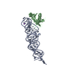 3ktvC 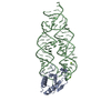 1lngS C: citing same article ( S: Starting model for refinement |
|---|---|
| Similar structure data |

-Links
 PDBj
PDBj


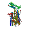
- Assembly
| Deposited unit | 
| ||||||||
|---|---|---|---|---|---|---|---|---|---|
| 1 | 
| ||||||||
| 2 | 
| ||||||||
| Unit cell |
|
-Components
| #1: RNA chain | Mass: 31165.553 Da / Num. of mol.: 2 / Fragment: S domain Source method: isolated from a genetically manipulated source Details: in vitro transcription / Source: (gene. exp.) Sulfolobus solfataricus (archaea) / Plasmid: pUC19 / Production host:  #2: Protein | Mass: 12939.142 Da / Num. of mol.: 2 Source method: isolated from a genetically manipulated source Source: (gene. exp.) Sulfolobus solfataricus (archaea) / Gene: srp19, SSO0165 / Plasmid: pET24d / Production host: #3: Chemical | ChemComp-K /   Mass: 39.098 Da / Num. of mol.: 4 / Source method: obtained synthetically / Formula: K Mass: 39.098 Da / Num. of mol.: 4 / Source method: obtained synthetically / Formula: K#4: Chemical | ChemComp-MG /   Mass: 24.305 Da / Num. of mol.: 6 / Source method: obtained synthetically / Formula: Mg Mass: 24.305 Da / Num. of mol.: 6 / Source method: obtained synthetically / Formula: Mg |
|---|
-Experimental details
-Experiment
| Experiment | Method: X-RAY DIFFRACTION / Number of used crystals: 1 |
|---|
- Sample preparation
| Crystal | Density Matthews: 3.58 Å3/Da / Density % sol: 65.62 % | ||||||||||||||||||||||||||||||||||||
|---|---|---|---|---|---|---|---|---|---|---|---|---|---|---|---|---|---|---|---|---|---|---|---|---|---|---|---|---|---|---|---|---|---|---|---|---|---|
| Crystal grow | Temperature: 293 K / Method: vapor diffusion, hanging drop / pH: 5.7 Details: 100 mM Na cacodylate, 400 mM KCl, 10 mM CaCl2, 15% (w/v) PEG4000, pH 5.7, VAPOR DIFFUSION, HANGING DROP, temperature 293K | ||||||||||||||||||||||||||||||||||||
| Components of the solutions |
|
-Data collection
| Diffraction | Mean temperature: 100 K |
|---|---|
| Diffraction source | Source: SYNCHROTRON / Site: ESRF  / Beamline: ID14-2 / Wavelength: 0.933 Å / Beamline: ID14-2 / Wavelength: 0.933 Å |
| Detector | Type: ADSC QUANTUM 4 / Detector: CCD / Date: Jun 23, 2006 / Details: mirrors |
| Radiation | Monochromator: Diamond (111), Ge (220) / Protocol: SINGLE WAVELENGTH / Monochromatic (M) / Laue (L): M / Scattering type: x-ray |
| Radiation wavelength | Wavelength: 0.933 Å / Relative weight: 1 |
| Reflection | Resolution: 3.2→64.7 Å / Num. all: 20744 / Num. obs: 20578 / % possible obs: 99.2 % / Observed criterion σ(F): 0 / Observed criterion σ(I): 0 / Redundancy: 3.9 % / Rsym value: 0.092 / Net I/σ(I): 13.6 |
| Reflection shell | Resolution: 3.2→3.37 Å / Redundancy: 3.9 % / Mean I/σ(I) obs: 3.3 / Rsym value: 0.388 / % possible all: 99.2 |
- Processing
| Software |
| |||||||||||||||||||||||||||||||||||||||||||||||||||||||||||||||||
|---|---|---|---|---|---|---|---|---|---|---|---|---|---|---|---|---|---|---|---|---|---|---|---|---|---|---|---|---|---|---|---|---|---|---|---|---|---|---|---|---|---|---|---|---|---|---|---|---|---|---|---|---|---|---|---|---|---|---|---|---|---|---|---|---|---|---|
| Refinement | Method to determine structure: MOLECULAR REPLACEMENT Starting model: PDB entry 1LNG Resolution: 3.2→64.68 Å / Cor.coef. Fo:Fc: 0.915 / Cor.coef. Fo:Fc free: 0.896 / Occupancy max: 1 / Occupancy min: 1 / SU B: 23.527 / SU ML: 0.412 / Cross valid method: THROUGHOUT / σ(F): 0 / σ(I): 0 / ESU R Free: 0.501 / Stereochemistry target values: MAXIMUM LIKELIHOOD Details: HYDROGENS HAVE BEEN ADDED IN THE RIDING POSITIONS. U VALUES REFINED INDIVIDUALLY
| |||||||||||||||||||||||||||||||||||||||||||||||||||||||||||||||||
| Solvent computation | Ion probe radii: 0.8 Å / Shrinkage radii: 0.8 Å / VDW probe radii: 1.2 Å / Solvent model: MASK | |||||||||||||||||||||||||||||||||||||||||||||||||||||||||||||||||
| Displacement parameters | Biso max: 184.16 Å2 / Biso mean: 86.721 Å2 / Biso min: 32.73 Å2
| |||||||||||||||||||||||||||||||||||||||||||||||||||||||||||||||||
| Refinement step | Cycle: LAST / Resolution: 3.2→64.68 Å
| |||||||||||||||||||||||||||||||||||||||||||||||||||||||||||||||||
| Refine LS restraints |
| |||||||||||||||||||||||||||||||||||||||||||||||||||||||||||||||||
| LS refinement shell | Resolution: 3.2→3.283 Å / Total num. of bins used: 20
|
