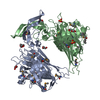
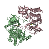
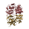
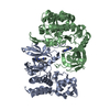
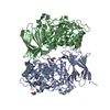
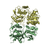
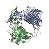
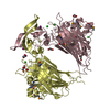
- PDB-3ks7: Crystal structure of Putative Peptide:N-glycosidase F (PNGase F) ... -
+
Open data
ID or keywords:
Loading...
-
Basic information
Entry
Database: PDB / ID: 3ks7
Title
Crystal structure of Putative Peptide:N-glycosidase F (PNGase F) (YP_210507.1) from Bacteroides fragilis NCTC 9343 at 2.30 A resolution
Components
Putative Putative PNGase F
Keywords
HYDROLASE / Putative Peptide:N-glycosidase F (PNGase F) / Structural Genomics / Joint Center for Structural Genomics / JCSG / Protein Structure Initiative / PSI-2
Function / homology
Function and homology information
oxidoreductase activity, acting on paired donors, with incorporation or reduction of molecular oxygen, reduced ascorbate as one donor, and incorporation of one atom of oxygen Similarity search - Function
Peptide-N-glycosidase F, N-terminal domain / Peptide-N-glycosidase F, N-terminal domain superfamily / Peptide-N-glycosidase F, N-terminal / Peptide-N-glycosidase F, N terminal / Peptide-N-glycosidase F, C-terminal / Peptide-N-glycosidase F, N terminal / Peptide-N-glycosidase F, C terminal / Jelly Rolls - #230 / PHM/PNGase F domain superfamily / Copper type II, ascorbate-dependent monooxygenase-like, C-terminal ...Peptide-N-glycosidase F, N-terminal domain / Peptide-N-glycosidase F, N-terminal domain superfamily / Peptide-N-glycosidase F, N-terminal / Peptide-N-glycosidase F, N terminal / Peptide-N-glycosidase F, C-terminal / Peptide-N-glycosidase F, N terminal / Peptide-N-glycosidase F, C terminal / Jelly Rolls - #230 / PHM/PNGase F domain superfamily / Copper type II, ascorbate-dependent monooxygenase-like, C-terminal / Jelly Rolls / Sandwich / Mainly Beta Similarity search - Domain/homology
ANALYTICAL SIZE EXCULSION CHROMATOGRAPHY WITH STATIC LIGHT SCATTERING SUPPORTS THE ASSIGNMENT OF THE DIMER AS A SIGNIFICANT OLIGOMERIZATION STATE IN SOLUTION.
-
Components
#1: Protein
PutativePutativePNGaseF
Mass: 44731.625 Da / Num. of mol.: 4 Source method: isolated from a genetically manipulated source Source: (gene. exp.) Bacteroides fragilis NCTC 9343 (bacteria) Strain: ATCC 25285 / NCTC 9343 / Gene: BF0811 / Plasmid: SpeedET / Production host: Escherichia Coli (E. coli) / Strain (production host): HK100 / References: UniProt: Q5LH31
Mass: 18.015 Da / Num. of mol.: 456 / Source method: isolated from a natural source / Formula: H2O
Has protein modification
Y
Sequence details
SEQUENCE: THIS CONSTRUCT (RESIDUE 20-415) WAS EXPRESSED WITH A PURIFICATION TAG MGSDKIHHHHHHENLYFQG. ...SEQUENCE: THIS CONSTRUCT (RESIDUE 20-415) WAS EXPRESSED WITH A PURIFICATION TAG MGSDKIHHHHHHENLYFQG. THE TAG WAS REMOVED WITH TEV PROTEASE LEAVING ONLY A GLYCINE (0) FOLLOWED BY THE TARGET SEQUENCE.
-
Experimental details
-
Experiment
Experiment
Method: X-RAY DIFFRACTION / Number of used crystals: 1
-
Sample preparation
Crystal
Density Matthews: 2.73 Å3/Da / Density % sol: 54.88 %
Crystal grow
Temperature: 277 K / Method: vapor diffusion, sitting drop Details: 25.0000% polyethylene glycol 3350, 0.2070M ammonium iodide, NANODROP, VAPOR DIFFUSION, SITTING DROP, temperature 277K
Monochromator: Single crystal Si(111) bent monochromator (horizontal focusing) Protocol: MAD / Monochromatic (M) / Laue (L): M / Scattering type: x-ray
Radiation wavelength
ID
Wavelength (Å)
Relative weight
1
0.91837
1
2
0.97922
1
3
0.97876
1
Reflection
Resolution: 2.3→29.881 Å / Num. obs: 87284 / % possible obs: 99.8 % / Redundancy: 3.7 % / Biso Wilson estimate: 43.391 Å2 / Rmerge(I) obs: 0.099 / Rsym value: 0.099 / Net I/σ(I): 9.9
Reflection shell
Diffraction-ID: 1
Resolution (Å)
Redundancy (%)
Rmerge(I) obs
Mean I/σ(I) obs
Num. measured all
Num. unique all
Rsym value
% possible all
2.3-2.36
3.8
0.789
1
24125
6398
0.789
99.9
2.36-2.42
3.8
0.702
1.1
23376
6227
0.702
100
2.42-2.49
3.8
0.569
1.3
22829
6071
0.569
99.9
2.49-2.57
3.8
0.486
1.6
22089
5887
0.486
100
2.57-2.66
3.8
0.373
2.1
21570
5732
0.373
99.9
2.66-2.75
3.8
0.304
2.5
20826
5532
0.304
100
2.75-2.85
3.8
0.249
3.1
20014
5327
0.249
100
2.85-2.97
3.8
0.196
3.9
19440
5172
0.196
100
2.97-3.1
3.8
0.16
4.7
18540
4934
0.16
100
3.1-3.25
3.8
0.117
6.4
17820
4746
0.117
100
3.25-3.43
3.7
0.086
8.5
16972
4533
0.086
100
3.43-3.64
3.8
0.068
10.2
15996
4260
0.068
100
3.64-3.89
3.7
0.063
10.8
15156
4046
0.063
99.9
3.89-4.2
3.7
0.059
10.8
13960
3747
0.059
99.9
4.2-4.6
3.7
0.05
12.2
12919
3473
0.05
99.8
4.6-5.14
3.7
0.046
13.6
11739
3155
0.046
99.8
5.14-5.94
3.7
0.049
13.4
10324
2792
0.049
99.6
5.94-7.27
3.7
0.05
13.4
8707
2370
0.05
99.6
7.27-10.29
3.6
0.045
13.6
6730
1862
0.045
99.2
10.29-29.88
3.4
0.041
11.5
3481
1020
0.041
93.9
-
Phasing
Phasing
Method: MAD
-
Processing
Software
Name
Version
Classification
NB
REFMAC
5.5.0102
refinement
PHENIX
refinement
SHELX
phasing
MolProbity
3beta29
modelbuilding
SCALA
3.2.5
datascaling
PDB_EXTRACT
3.006
dataextraction
MOSFLM
datareduction
SHELXD
phasing
autoSHARP
phasing
Refinement
Method to determine structure: MAD / Resolution: 2.3→29.881 Å / Cor.coef. Fo:Fc: 0.948 / Cor.coef. Fo:Fc free: 0.929 / Occupancy max: 1 / Occupancy min: 0.33 / SU B: 15.772 / SU ML: 0.17 / TLS residual ADP flag: LIKELY RESIDUAL / Cross valid method: THROUGHOUT / σ(F): 0 / ESU R: 0.302 / ESU R Free: 0.219 Stereochemistry target values: MAXIMUM LIKELIHOOD WITH PHASES Details: 1. HYDROGENS HAVE BEEN ADDED IN THE RIDING POSITIONS. 2. A MET-INHIBITION PROTOCOL WAS USED FOR SELENOMETHIONINE INCORPORATION DURING PROTEIN EXPRESSION. THE OCCUPANCY OF THE SE ATOMS IN THE ...Details: 1. HYDROGENS HAVE BEEN ADDED IN THE RIDING POSITIONS. 2. A MET-INHIBITION PROTOCOL WAS USED FOR SELENOMETHIONINE INCORPORATION DURING PROTEIN EXPRESSION. THE OCCUPANCY OF THE SE ATOMS IN THE MSE RESIDUES WAS REDUCED TO 0.75 FOR THE REDUCED SCATTERING POWER DUE TO PARTIAL S-MET INCORPORATION. 3. TLS GROUPS WERE ASSIGNED WITH THE AID OF THE TLS MOTION DETERMINATION SERVER. 4. ANOMALOUS DIFFERENCE MAPS SUPPORTED THE MODELING OF IODIDE (IOD) FROM THE CRYSTALLIZATION SOLUTION. THE OCCUPANCIES ON THE IODIDES WERE REDUCED TO ACCOUNT FOR THE OBSERVED SCATTERING. 5. ETHYLENE GLYCOL (EDO) USED AS A CRYOPROTECTANT WAS MODELED INTO THE STRUCTURE AND CHLORIDE (CL) FROM THE PURIFICATION/ CRYSTALLIZATION SOLUTIONS WERE MODELED INTO THE STRUCTURE.
Rfactor
Num. reflection
% reflection
Selection details
Rfree
0.237
4375
5 %
RANDOM
Rwork
0.201
-
-
-
obs
0.203
87221
99.74 %
-
Solvent computation
Ion probe radii: 0.8 Å / Shrinkage radii: 0.8 Å / VDW probe radii: 1.4 Å / Solvent model: MASK
In the structure databanks used in Yorodumi, some data are registered as the other names, "COVID-19 virus" and "2019-nCoV". Here are the details of the virus and the list of structure data.
Jan 31, 2019. EMDB accession codes are about to change! (news from PDBe EMDB page)
EMDB accession codes are about to change! (news from PDBe EMDB page)
The allocation of 4 digits for EMDB accession codes will soon come to an end. Whilst these codes will remain in use, new EMDB accession codes will include an additional digit and will expand incrementally as the available range of codes is exhausted. The current 4-digit format prefixed with “EMD-” (i.e. EMD-XXXX) will advance to a 5-digit format (i.e. EMD-XXXXX), and so on. It is currently estimated that the 4-digit codes will be depleted around Spring 2019, at which point the 5-digit format will come into force.
The EM Navigator/Yorodumi systems omit the EMD- prefix.
Related info.:Q: What is EMD? / ID/Accession-code notation in Yorodumi/EM Navigator
Yorodumi is a browser for structure data from EMDB, PDB, SASBDB, etc.
This page is also the successor to EM Navigator detail page, and also detail information page/front-end page for Omokage search.
The word "yorodu" (or yorozu) is an old Japanese word meaning "ten thousand". "mi" (miru) is to see.
Related info.:EMDB / PDB / SASBDB / Comparison of 3 databanks / Yorodumi Search / Aug 31, 2016. New EM Navigator & Yorodumi / Yorodumi Papers / Jmol/JSmol / Function and homology information / Changes in new EM Navigator and Yorodumi
 Movie
Movie Controller

 Yorodumi
 Open data
 Basic information
 Components
 Keywords
 Function and homology information
 Bacteroides fragilis NCTC 9343 (bacteria)
 X-RAY DIFFRACTION /
  Authors
 Citation
 Structure visualization
 Downloads & links
 Other downloads
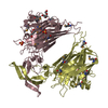

 PDBj
PDBj Assembly
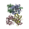

 Mass: 126.904 Da / Num. of mol.: 7 / Source method: obtained synthetically / Formula: I
 Mass: 35.453 Da / Num. of mol.: 10 / Source method: obtained synthetically / Formula: Cl
 Mass: 62.068 Da / Num. of mol.: 22 / Source method: obtained synthetically / Formula: C2H6O2
 Mass: 18.015 Da / Num. of mol.: 456 / Source method: isolated from a natural source / Formula: H2O
 Sample preparation
 / Beamline: BL11-1 / Wavelength: 0.91837,0.97922,0.97876
 Processing