+ Open data
- Basic information
| Entry | Database: PDB / ID: 3kqa | ||||||
|---|---|---|---|---|---|---|---|
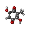
| Title | MurA dead-end complex with terreic acid | ||||||
 Components Components | UDP-N-acetylglucosamine 1-carboxyvinyltransferase | ||||||
 Keywords Keywords | TRANSFERASE/TRANSFERASE INHIBITOR / Open enzyme state / inside-out alpha/beta barrel / Cell cycle / Cell division / Cell shape / Cell wall biogenesis/degradation / Peptidoglycan synthesis / TRANSFERASE / TRANSFERASE-TRANSFERASE INHIBITOR complex | ||||||
| Function / homology |  Function and homology information Function and homology informationUDP-N-acetylglucosamine 1-carboxyvinyltransferase activity / UDP-N-acetylgalactosamine biosynthetic process / UDP-N-acetylglucosamine 1-carboxyvinyltransferase / peptidoglycan biosynthetic process / cell wall organization / regulation of cell shape / cell division / cytoplasm Similarity search - Function | ||||||
| Biological species |  Enterobacter cloacae (bacteria) Enterobacter cloacae (bacteria) | ||||||
| Method |  X-RAY DIFFRACTION / X-RAY DIFFRACTION /  MOLECULAR REPLACEMENT / Resolution: 2.25 Å MOLECULAR REPLACEMENT / Resolution: 2.25 Å | ||||||
 Authors Authors | Schonbrunn, E. | ||||||
 Citation Citation |  Journal: Biochemistry / Year: 2010 Journal: Biochemistry / Year: 2010Title: The fungal product terreic acid is a covalent inhibitor of the bacterial cell wall biosynthetic enzyme UDP-N-acetylglucosamine 1-carboxyvinyltransferase (MurA) . Authors: Han, H. / Yang, Y. / Olesen, S.H. / Becker, A. / Betzi, S. / Schonbrunn, E. | ||||||
| History |
|
- Structure visualization
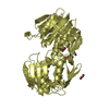
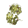
| Structure viewer | Molecule:  Molmil Molmil Jmol/JSmol Jmol/JSmol |
|---|
- Downloads & links
- Download
| PDBx/mmCIF format |  3kqa.cif.gz 3kqa.cif.gz | 327.9 KB | Display |  PDBx/mmCIF format PDBx/mmCIF format |
|---|---|---|---|---|
| PDB format |  pdb3kqa.ent.gz pdb3kqa.ent.gz | 267.5 KB | Display |  PDB format PDB format |
| PDBx/mmJSON format |  3kqa.json.gz 3kqa.json.gz | Tree view |  PDBx/mmJSON format PDBx/mmJSON format | |
| Others |  Other downloads Other downloads |
-Validation report
| Arichive directory |  https://data.pdbj.org/pub/pdb/validation_reports/kq/3kqa https://data.pdbj.org/pub/pdb/validation_reports/kq/3kqa ftp://data.pdbj.org/pub/pdb/validation_reports/kq/3kqa ftp://data.pdbj.org/pub/pdb/validation_reports/kq/3kqa | HTTPS FTP |
|---|
-Related structure data
| Related structure data |  3kr6C  3lthC 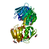 1ejcS C: citing same article ( S: Starting model for refinement |
|---|---|
| Similar structure data |
- Links
Links
- Assembly
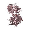
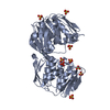
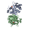
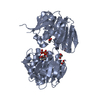
| Deposited unit | 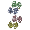
| |||||||||
|---|---|---|---|---|---|---|---|---|---|---|
| 1 | 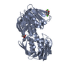
| |||||||||
| 2 | 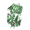
| |||||||||
| 3 | 
| |||||||||
| 4 | 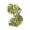
| |||||||||
| Unit cell |
| |||||||||
| Components on special symmetry positions |
|
- Components
| #1: Protein | Mass: 44829.406 Da / Num. of mol.: 4 Source method: isolated from a genetically manipulated source Source: (gene. exp.)  Enterobacter cloacae (bacteria) / Gene: murA, MurA (MurZ), murZ / Plasmid: pET9d / Production host: Enterobacter cloacae (bacteria) / Gene: murA, MurA (MurZ), murZ / Plasmid: pET9d / Production host:  References: UniProt: P33038, UDP-N-acetylglucosamine 1-carboxyvinyltransferase #2: Chemical | ChemComp-TR9 / ( #3: Chemical | ChemComp-CA / #4: Water | ChemComp-HOH / | Has protein modification | Y | Nonpolymer details | THE UNBOUND FROM OF THE INHIBITOR TR9 IS TERREIC ACID. UPON REACTION WITH PROTEIN IT BINDS ...THE UNBOUND FROM OF THE INHIBITOR TR9 IS TERREIC ACID. UPON REACTION WITH PROTEIN IT BINDS COVALENTLY | Sequence details | ASP67 FORMS AN ISOPEPTIDI | |
|---|
-Experimental details
-Experiment
| Experiment | Method:  X-RAY DIFFRACTION / Number of used crystals: 1 X-RAY DIFFRACTION / Number of used crystals: 1 |
|---|
- Sample preparation
| Crystal | Density Matthews: 2.56 Å3/Da / Density % sol: 51.96 % |
|---|---|
| Crystal grow | Temperature: 292 K / Method: vapor diffusion, hanging drop / pH: 7.5 Details: 100 mM CaCl2, 100 mM HEPES, pH 7.5, 30% PEG 400, VAPOR DIFFUSION, HANGING DROP, temperature 292K |
-Data collection
| Diffraction | Mean temperature: 93 K |
|---|---|
| Diffraction source | Source:  ROTATING ANODE / Type: RIGAKU MICROMAX-007 HF / Wavelength: 1.542 Å ROTATING ANODE / Type: RIGAKU MICROMAX-007 HF / Wavelength: 1.542 Å |
| Detector | Type: RIGAKU RAXIS HTC / Detector: IMAGE PLATE / Date: Jan 15, 2009 / Details: mirrors |
| Radiation | Monochromator: mirrors / Protocol: SINGLE WAVELENGTH / Monochromatic (M) / Laue (L): M / Scattering type: x-ray |
| Radiation wavelength | Wavelength: 1.542 Å / Relative weight: 1 |
| Reflection | Resolution: 2.25→15 Å / Num. all: 88863 / Num. obs: 88863 / % possible obs: 99.4 % / Observed criterion σ(F): 0 / Observed criterion σ(I): -3 / Redundancy: 5.2 % / Biso Wilson estimate: 22.1 Å2 / Rmerge(I) obs: 0.049 / Net I/σ(I): 23.4 |
| Reflection shell | Resolution: 2.25→2.3 Å / Redundancy: 3 % / Rmerge(I) obs: 0.175 / Mean I/σ(I) obs: 7.9 / Num. unique all: 5612 / Rsym value: 0.079 / % possible all: 100 |
- Processing
| Software |
| |||||||||||||||||||||||||
|---|---|---|---|---|---|---|---|---|---|---|---|---|---|---|---|---|---|---|---|---|---|---|---|---|---|---|
| Refinement | Method to determine structure:  MOLECULAR REPLACEMENT MOLECULAR REPLACEMENTStarting model: 1EJC Resolution: 2.25→15 Å / Cross valid method: THROUGHOUT / σ(F): 0 / σ(I): -3 / Stereochemistry target values: Engh & Huber
| |||||||||||||||||||||||||
| Refine analyze |
| |||||||||||||||||||||||||
| Refinement step | Cycle: LAST / Resolution: 2.25→15 Å
| |||||||||||||||||||||||||
| LS refinement shell | Resolution: 2.25→2.39 Å / Rfactor Rfree error: 0.023
|
 Movie
Movie Controller


 PDBj
PDBj

