- PDB-3kop: Crystal structure of Protein with a cyclophilin-like fold (YP_831... -
+
Open data
ID or keywords:
Loading...
-
Basic information
Entry
Database: PDB / ID: 3kop
Title
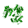
Crystal structure of Protein with a cyclophilin-like fold (YP_831253.1) from Arthrobacter sp. FB24 at 1.90 A resolution
Components
Uncharacterized protein
Keywords
Structural Genomics / Unknown function / Protein with a cyclophilin-like fold / Joint Center for Structural Genomics / JCSG / Protein Structure Initiative / PSI-2
Function / homology
Protein of unknown function DUF3830 / Protein of unknown function (DUF3830) / Cyclophilin - #20 / Cyclophilin / Cyclophilin-like domain superfamily / Beta Barrel / Mainly Beta / DUF3830 domain-containing protein
Function and homology information
Biological species
Arthrobacter sp. (bacteria)
Method
X-RAY DIFFRACTION / SYNCHROTRON / MAD / Resolution: 1.9 Å
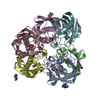
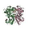
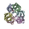
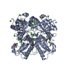
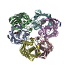
A: Uncharacterized protein B: Uncharacterized protein C: Uncharacterized protein D: Uncharacterized protein E: Uncharacterized protein F: Uncharacterized protein
Monochromator: Single crystal Si(111) bent monochromator (horizontal focusing) Protocol: MAD / Monochromatic (M) / Laue (L): M / Scattering type: x-ray
Radiation wavelength
ID
Wavelength (Å)
Relative weight
1
0.91837
1
2
0.97963
1
Reflection
Resolution: 1.9→29.907 Å / Num. obs: 88263 / % possible obs: 99.5 % / Redundancy: 2.6 % / Biso Wilson estimate: 18.599 Å2 / Rmerge(I) obs: 0.086 / Rsym value: 0.086 / Net I/σ(I): 9
Reflection shell
Diffraction-ID: 1
Resolution (Å)
Redundancy (%)
Rmerge(I) obs
Mean I/σ(I) obs
Num. measured all
Num. unique all
Rsym value
% possible all
1.9-1.95
2.5
0.498
2
16367
6518
0.498
100
1.95-2
2.5
0.399
1.9
16215
6414
0.399
99.9
2-2.06
2.5
0.313
2.4
15622
6195
0.313
100
2.06-2.12
2.5
0.302
0.9
15339
6075
0.302
100
2.12-2.19
2.5
0.224
3.4
14709
5797
0.224
99.9
2.19-2.27
2.5
0.215
1.3
14349
5664
0.215
99.9
2.27-2.36
2.5
0.173
4.3
13863
5442
0.173
99.9
2.36-2.45
2.6
0.143
5.2
13407
5245
0.143
99.8
2.45-2.56
2.6
0.126
5.9
12792
5008
0.126
99.7
2.56-2.69
2.6
0.111
6.2
12198
4782
0.111
99.6
2.69-2.83
2.6
0.089
8
11708
4564
0.089
99.5
2.83-3
2.6
0.08
8.5
11149
4330
0.08
99.4
3-3.21
2.6
0.071
8.9
10343
4029
0.071
99.2
3.21-3.47
2.6
0.057
10.5
9722
3773
0.057
99.2
3.47-3.8
2.6
0.047
12.8
8962
3462
0.047
98.8
3.8-4.25
2.6
0.041
12.6
8129
3136
0.041
98.6
4.25-4.91
2.6
0.036
16.2
7106
2731
0.036
98.2
4.91-6.01
2.6
0.037
16.2
6067
2334
0.037
98
6.01-8.5
2.6
0.045
13
4682
1797
0.045
97.5
8.5-29.91
2.6
0.041
12.3
2479
967
0.041
94.4
-
Phasing
Phasing
Method: MAD
-
Processing
Software
Name
Version
Classification
NB
REFMAC
5.5.0053
refinement
PHENIX
refinement
SOLVE
phasing
MolProbity
3beta29
modelbuilding
SCALA
3.2.5
datascaling
PDB_EXTRACT
3.006
dataextraction
MOSFLM
datareduction
Refinement
Method to determine structure: MAD / Resolution: 1.9→29.907 Å / Cor.coef. Fo:Fc: 0.96 / Cor.coef. Fo:Fc free: 0.939 / Occupancy max: 1 / Occupancy min: 0.3 / SU B: 6.209 / SU ML: 0.08 / TLS residual ADP flag: LIKELY RESIDUAL / Cross valid method: THROUGHOUT / σ(F): 0 / ESU R: 0.128 / ESU R Free: 0.121 Stereochemistry target values: MAXIMUM LIKELIHOOD WITH PHASES Details: 1. HYDROGENS HAVE BEEN ADDED IN THE RIDING POSITIONS. 2. A MET-INHIBITION PROTOCOL WAS USED FOR SELENOMETHIONINE INCORPORATION DURING PROTEIN EXPRESSION. THE OCCUPANCY OF THE SE ATOMS IN THE ...Details: 1. HYDROGENS HAVE BEEN ADDED IN THE RIDING POSITIONS. 2. A MET-INHIBITION PROTOCOL WAS USED FOR SELENOMETHIONINE INCORPORATION DURING PROTEIN EXPRESSION. THE OCCUPANCY OF THE SE ATOMS IN THE MSE RESIDUES WAS REDUCED TO 0.75 FOR THE REDUCED SCATTERING POWER DUE TO PARTIAL S-MET INCORPORATION. 3. ATOM RECORDS CONTAIN RESIDUAL B FACTORS ONLY.
Rfactor
Num. reflection
% reflection
Selection details
Rfree
0.196
4416
5 %
RANDOM
Rwork
0.16
-
-
-
obs
0.162
88235
99.3 %
-
Solvent computation
Ion probe radii: 0.8 Å / Shrinkage radii: 0.8 Å / VDW probe radii: 1.2 Å / Solvent model: MASK
In the structure databanks used in Yorodumi, some data are registered as the other names, "COVID-19 virus" and "2019-nCoV". Here are the details of the virus and the list of structure data.
Jan 31, 2019. EMDB accession codes are about to change! (news from PDBe EMDB page)
EMDB accession codes are about to change! (news from PDBe EMDB page)
The allocation of 4 digits for EMDB accession codes will soon come to an end. Whilst these codes will remain in use, new EMDB accession codes will include an additional digit and will expand incrementally as the available range of codes is exhausted. The current 4-digit format prefixed with “EMD-” (i.e. EMD-XXXX) will advance to a 5-digit format (i.e. EMD-XXXXX), and so on. It is currently estimated that the 4-digit codes will be depleted around Spring 2019, at which point the 5-digit format will come into force.
The EM Navigator/Yorodumi systems omit the EMD- prefix.
Related info.:Q: What is EMD? / ID/Accession-code notation in Yorodumi/EM Navigator
Yorodumi is a browser for structure data from EMDB, PDB, SASBDB, etc.
This page is also the successor to EM Navigator detail page, and also detail information page/front-end page for Omokage search.
The word "yorodu" (or yorozu) is an old Japanese word meaning "ten thousand". "mi" (miru) is to see.
Related info.:EMDB / PDB / SASBDB / Comparison of 3 databanks / Yorodumi Search / Aug 31, 2016. New EM Navigator & Yorodumi / Yorodumi Papers / Jmol/JSmol / Function and homology information / Changes in new EM Navigator and Yorodumi
 Movie
Movie Controller

 Yorodumi
 Open data
 Basic information
 Components
 Keywords
 Function and homology information
 Arthrobacter sp. (bacteria)
 X-RAY DIFFRACTION /
  Authors
 Citation
 Structure visualization
 Downloads & links
 Other downloads
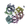
 PDBj
PDBj

 Assembly
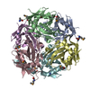
 Mass: 18.015 Da / Num. of mol.: 814 / Source method: isolated from a natural source / Formula: H2O
 Sample preparation
 / Beamline: BL11-1 / Wavelength: 0.91837,0.97963
 Processing