- PDB-3kkq: Crystal structure of M-Ras P40D in complex with GDP -
+
Open data
ID or keywords:
Loading...
-
Basic information
Entry
Database: PDB / ID: 3kkq
Title
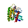
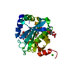
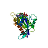
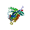
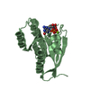
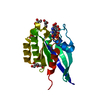
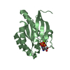
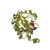
Crystal structure of M-Ras P40D in complex with GDP
Components
Ras-related protein M-Ras
Keywords
SIGNALING PROTEIN / GTP-binding / GTPase
Function / homology
Function and homology information
RAF activation / protein phosphatase type 1 complex / GTP-dependent protein binding / cellular response to leukemia inhibitory factor / small monomeric GTPase / GDP binding / G protein activity / Ras protein signal transduction / GTPase activity / GTP binding / plasma membrane Similarity search - Function
Small GTPase, Ras-type / Small GTPase Ras domain profile. / Ran (Ras-related nuclear proteins) /TC4 subfamily of small GTPases / Rho (Ras homology) subfamily of Ras-like small GTPases / Ras subfamily of RAS small GTPases / Small GTPase / Ras family / Rab subfamily of small GTPases / Small GTP-binding protein domain / P-loop containing nucleotide triphosphate hydrolases ...Small GTPase, Ras-type / Small GTPase Ras domain profile. / Ran (Ras-related nuclear proteins) /TC4 subfamily of small GTPases / Rho (Ras homology) subfamily of Ras-like small GTPases / Ras subfamily of RAS small GTPases / Small GTPase / Ras family / Rab subfamily of small GTPases / Small GTP-binding protein domain / P-loop containing nucleotide triphosphate hydrolases / Rossmann fold / P-loop containing nucleoside triphosphate hydrolase / 3-Layer(aba) Sandwich / Alpha Beta Similarity search - Domain/homology
Resolution: 1.2→29.51 Å / Cor.coef. Fo:Fc: 0.961 / Cor.coef. Fo:Fc free: 0.953 / SU B: 0.466 / SU ML: 0.022 / Cross valid method: THROUGHOUT / ESU R: 0.038 / ESU R Free: 0.04 / Stereochemistry target values: MAXIMUM LIKELIHOOD / Details: HYDROGENS HAVE BEEN ADDED IN THE RIDING POSITIONS
Rfactor
Num. reflection
% reflection
Selection details
Rfree
0.19222
3159
5.1 %
RANDOM
Rwork
0.17576
-
-
-
all
0.17658
59019
-
-
obs
0.17658
59019
98.18 %
-
Solvent computation
Ion probe radii: 0.8 Å / Shrinkage radii: 0.8 Å / VDW probe radii: 1.2 Å / Solvent model: MASK
Displacement parameters
Biso mean: 10.329 Å2
Baniso -1
Baniso -2
Baniso -3
1-
0 Å2
0 Å2
0 Å2
2-
-
0 Å2
0 Å2
3-
-
-
0 Å2
Refinement step
Cycle: LAST / Resolution: 1.2→29.51 Å
Protein
Nucleic acid
Ligand
Solvent
Total
Num. atoms
1387
0
29
243
1659
Refine LS restraints
Refine-ID
Type
Dev ideal
Dev ideal target
Number
X-RAY DIFFRACTION
r_bond_refined_d
0.006
0.022
1445
X-RAY DIFFRACTION
r_angle_refined_deg
1.161
1.982
1961
X-RAY DIFFRACTION
r_dihedral_angle_1_deg
5.425
5
169
X-RAY DIFFRACTION
r_dihedral_angle_2_deg
36.544
24.444
72
X-RAY DIFFRACTION
r_dihedral_angle_3_deg
11.436
15
256
X-RAY DIFFRACTION
r_dihedral_angle_4_deg
16.952
15
9
X-RAY DIFFRACTION
r_chiral_restr
0.077
0.2
218
X-RAY DIFFRACTION
r_gen_planes_refined
0.004
0.02
1084
X-RAY DIFFRACTION
r_nbd_refined
0.192
0.2
678
X-RAY DIFFRACTION
r_nbtor_refined
0.311
0.2
997
X-RAY DIFFRACTION
r_xyhbond_nbd_refined
0.097
0.2
185
X-RAY DIFFRACTION
r_symmetry_vdw_refined
0.155
0.2
35
X-RAY DIFFRACTION
r_symmetry_hbond_refined
0.088
0.2
22
X-RAY DIFFRACTION
r_mcbond_it
0.61
1.5
868
X-RAY DIFFRACTION
r_mcangle_it
0.991
2
1383
X-RAY DIFFRACTION
r_scbond_it
1.457
3
653
X-RAY DIFFRACTION
r_scangle_it
2.137
4.5
578
LS refinement shell
Resolution: 1.2→1.231 Å / Total num. of bins used: 20
Rfactor
Num. reflection
% reflection
Rfree
0.199
254
-
Rwork
0.19
4202
-
obs
-
4202
96.93 %
+
About Yorodumi
-
News
-
Feb 9, 2022. New format data for meta-information of EMDB entries
New format data for meta-information of EMDB entries
Version 3 of the EMDB header file is now the official format.
The previous official version 1.9 will be removed from the archive.
In the structure databanks used in Yorodumi, some data are registered as the other names, "COVID-19 virus" and "2019-nCoV". Here are the details of the virus and the list of structure data.
Jan 31, 2019. EMDB accession codes are about to change! (news from PDBe EMDB page)
EMDB accession codes are about to change! (news from PDBe EMDB page)
The allocation of 4 digits for EMDB accession codes will soon come to an end. Whilst these codes will remain in use, new EMDB accession codes will include an additional digit and will expand incrementally as the available range of codes is exhausted. The current 4-digit format prefixed with “EMD-” (i.e. EMD-XXXX) will advance to a 5-digit format (i.e. EMD-XXXXX), and so on. It is currently estimated that the 4-digit codes will be depleted around Spring 2019, at which point the 5-digit format will come into force.
The EM Navigator/Yorodumi systems omit the EMD- prefix.
Related info.:Q: What is EMD? / ID/Accession-code notation in Yorodumi/EM Navigator
Yorodumi is a browser for structure data from EMDB, PDB, SASBDB, etc.
This page is also the successor to EM Navigator detail page, and also detail information page/front-end page for Omokage search.
The word "yorodu" (or yorozu) is an old Japanese word meaning "ten thousand". "mi" (miru) is to see.
Related info.:EMDB / PDB / SASBDB / Comparison of 3 databanks / Yorodumi Search / Aug 31, 2016. New EM Navigator & Yorodumi / Yorodumi Papers / Jmol/JSmol / Function and homology information / Changes in new EM Navigator and Yorodumi
 Movie
Movie Controller


 Open data
 Basic information
 Components
 Keywords
 Function and homology information
 X-RAY DIFFRACTION /
  Authors
 Citation
 Structure visualization
 Downloads & links
 Other downloads
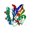


 PDBj
PDBj


 Assembly
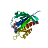


 Type: RNA linking / Mass: 443.201 Da / Num. of mol.: 1 / Source method: obtained synthetically / Formula: C10H15N5O11P2 / Comment: GDP, energy-carrying molecule*YM
 Mass: 24.305 Da / Num. of mol.: 1 / Source method: obtained synthetically / Formula: Mg
 Mass: 18.015 Da / Num. of mol.: 243 / Source method: isolated from a natural source / Formula: H2O
 Sample preparation
 / Beamline: BL41XU / Wavelength: 0.8 Å
 Processing