 Movie
Movie Controller

[English] 日本語
 Yorodumi
- PDB-3kkl: Crystal structure of functionally unknown HSP33 from Saccharomyce... -
+ Open data

- Basic information
| Entry | Database: PDB / ID: 3kkl | ||||||
|---|---|---|---|---|---|---|---|
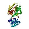
| Title | Crystal structure of functionally unknown HSP33 from Saccharomyces cerevisiae | ||||||
 Components Components | Probable chaperone protein HSP33 | ||||||
 Keywords Keywords | HYDROLASE / peptidase / heat shock protein / Chaperone / Protease / Stress response | ||||||
| Function / homology |  Function and homology information Function and homology informationD-lactate dehydratase / glyoxalase III activity / : / protein folding chaperone / cellular response to nutrient levels / P-body / protein folding / cytoplasm Similarity search - Function | ||||||
| Biological species |  | ||||||
| Method |  X-RAY DIFFRACTION / SYNCHROTRON / MOLECULAR REPLACEMENT / Resolution: 2.03 Å X-RAY DIFFRACTION / SYNCHROTRON / MOLECULAR REPLACEMENT / Resolution: 2.03 Å | ||||||
 Authors Authors | Hwang, K.Y. / Sung, M.W. / Lee, W.H. | ||||||
 Citation Citation | Journal: To be Published Title: Crystal structure of functionally unknown HSP33 from Saccharomyces cerevisiae Authors: Hwang, K.Y. / Sung, M.W. / Lee, W.H. | ||||||
| History |
|
- Structure visualization
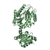
| Structure viewer | Molecule: MolmilJmol/JSmol |
|---|
- Downloads & links
-Download
| PDBx/mmCIF format | 3kkl.cif.gz | 93.6 KB | Display | PDBx/mmCIF format |
|---|---|---|---|---|
| PDB format | pdb3kkl.ent.gz | 71.8 KB | Display | PDB format |
| PDBx/mmJSON format | 3kkl.json.gz | Tree view | PDBx/mmJSON format | |
| Others |  Other downloads Other downloads |
-Validation report
| Arichive directory | https://data.pdbj.org/pub/pdb/validation_reports/kk/3kklftp://data.pdbj.org/pub/pdb/validation_reports/kk/3kkl | HTTPS FTP |
|---|
-Related structure data
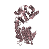
| Related structure data |  1qvzS S: Starting model for refinement |
|---|---|
| Similar structure data |


-Links
 PDBj
PDBj


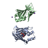
- Assembly
| Deposited unit | 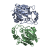
| ||||||||
|---|---|---|---|---|---|---|---|---|---|
| 1 |
| ||||||||
| Unit cell |
|
-Components
| #1: Protein | Mass: 26878.396 Da / Num. of mol.: 2 Source method: isolated from a genetically manipulated source Source: (gene. exp.) Gene: HSP33, YOR391C / Plasmid: pET22b / Production host:  #2: Water | ChemComp-HOH / |  Mass: 18.015 Da / Num. of mol.: 50 / Source method: isolated from a natural source / Formula: H2O Mass: 18.015 Da / Num. of mol.: 50 / Source method: isolated from a natural source / Formula: H2O |
|---|
-Experimental details
-Experiment
| Experiment | Method: X-RAY DIFFRACTION / Number of used crystals: 1 |
|---|
- Sample preparation
| Crystal | Density Matthews: 1.81 Å3/Da / Density % sol: 32.18 % |
|---|---|
| Crystal grow | Temperature: 295 K / Method: vapor diffusion, hanging drop / pH: 7.5 Details: 0.2M Tri-sodium Citrate dihydrate, 23%(w/v) polyethylene glycol 3350, pH 7.5, VAPOR DIFFUSION, HANGING DROP, temperature 295K |
-Data collection
| Diffraction | Mean temperature: 100 K |
|---|---|
| Diffraction source | Source: SYNCHROTRON / Site: PAL/PLS  / Beamline: 4A / Wavelength: 1 Å / Beamline: 4A / Wavelength: 1 Å |
| Detector | Type: ADSC QUANTUM 210 / Detector: CCD / Date: Sep 30, 2007 |
| Radiation | Monochromator: GRAPHITE / Protocol: SINGLE WAVELENGTH / Monochromatic (M) / Laue (L): M / Scattering type: x-ray |
| Radiation wavelength | Wavelength: 1 Å / Relative weight: 1 |
| Reflection | Resolution: 2.03→50 Å / Num. obs: 24915 / % possible obs: 92.5 % / Observed criterion σ(F): 2 / Observed criterion σ(I): 1 / Num. measured all: 109787 |
| Reflection shell | Resolution: 2.03→2.1 Å / % possible all: 87.5 |
- Processing
| Software |
| |||||||||||||||||||||||||||
|---|---|---|---|---|---|---|---|---|---|---|---|---|---|---|---|---|---|---|---|---|---|---|---|---|---|---|---|---|
| Refinement | Method to determine structure: MOLECULAR REPLACEMENT Starting model: PDB ENTRY 1qvz Resolution: 2.03→19.76 Å / Isotropic thermal model: RESTRAINED / Cross valid method: THROUGHOUT / σ(F): 0 / Stereochemistry target values: Engh & Huber
| |||||||||||||||||||||||||||
| Displacement parameters | Biso mean: 38.2 Å2
| |||||||||||||||||||||||||||
| Refine analyze |
| |||||||||||||||||||||||||||
| Refinement step | Cycle: LAST / Resolution: 2.03→19.76 Å
| |||||||||||||||||||||||||||
| Refine LS restraints |
|
