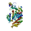
Unblocking of NMDA receptors, glutamate binding and activation / Ion transport by P-type ATPases / Ion homeostasis / Ca2+ pathway / HSF1-dependent transactivation / medium-term memory / serotonin biosynthetic process / Ca2+/calmodulin-dependent protein kinase / calcium/calmodulin-dependent protein kinase activity / regulation of neuronal synaptic plasticity ...Unblocking of NMDA receptors, glutamate binding and activation / Ion transport by P-type ATPases / Ion homeostasis / Ca2+ pathway / HSF1-dependent transactivation / medium-term memory / serotonin biosynthetic process / Ca2+/calmodulin-dependent protein kinase / calcium/calmodulin-dependent protein kinase activity / regulation of neuronal synaptic plasticity / regulation of protein localization to plasma membrane / axon cytoplasm / MAPK cascade / perikaryon / transmembrane transporter binding / calmodulin binding / neuron projection / postsynaptic density / protein serine kinase activity / positive regulation of gene expression / ATP binding / metal ion binding / identical protein binding / cytoplasm Similarity search - Function
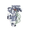
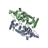
Calcium/calmodulin-dependent protein kinase II, association-domain / Calcium/calmodulin dependent protein kinase II association domain / NTF2-like domain superfamily / Phosphorylase Kinase; domain 1 / Phosphorylase Kinase; domain 1 / Transferase(Phosphotransferase) domain 1 / Transferase(Phosphotransferase); domain 1 / Serine/threonine-protein kinase, active site / Serine/Threonine protein kinases active-site signature. / Protein kinase domain ...Calcium/calmodulin-dependent protein kinase II, association-domain / Calcium/calmodulin dependent protein kinase II association domain / NTF2-like domain superfamily / Phosphorylase Kinase; domain 1 / Phosphorylase Kinase; domain 1 / Transferase(Phosphotransferase) domain 1 / Transferase(Phosphotransferase); domain 1 / Serine/threonine-protein kinase, active site / Serine/Threonine protein kinases active-site signature. / Protein kinase domain / Serine/Threonine protein kinases, catalytic domain / Protein kinase, ATP binding site / Protein kinases ATP-binding region signature. / Protein kinase domain profile. / Protein kinase domain / Protein kinase-like domain superfamily / 2-Layer Sandwich / Orthogonal Bundle / Mainly Alpha / Alpha Beta Similarity search - Domain/homology
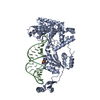
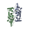
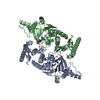
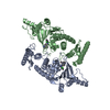
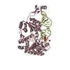
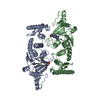
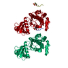
Calcium/calmodulin-dependent protein kinase type II / Calcium/calmodulin-dependent protein kinase type II Similarity search - Component
In the structure databanks used in Yorodumi, some data are registered as the other names, "COVID-19 virus" and "2019-nCoV". Here are the details of the virus and the list of structure data.
Jan 31, 2019. EMDB accession codes are about to change! (news from PDBe EMDB page)
EMDB accession codes are about to change! (news from PDBe EMDB page)
The allocation of 4 digits for EMDB accession codes will soon come to an end. Whilst these codes will remain in use, new EMDB accession codes will include an additional digit and will expand incrementally as the available range of codes is exhausted. The current 4-digit format prefixed with “EMD-” (i.e. EMD-XXXX) will advance to a 5-digit format (i.e. EMD-XXXXX), and so on. It is currently estimated that the 4-digit codes will be depleted around Spring 2019, at which point the 5-digit format will come into force.
The EM Navigator/Yorodumi systems omit the EMD- prefix.
Related info.:Q: What is EMD? / ID/Accession-code notation in Yorodumi/EM Navigator
Yorodumi is a browser for structure data from EMDB, PDB, SASBDB, etc.
This page is also the successor to EM Navigator detail page, and also detail information page/front-end page for Omokage search.
The word "yorodu" (or yorozu) is an old Japanese word meaning "ten thousand". "mi" (miru) is to see.
Related info.:EMDB / PDB / SASBDB / Comparison of 3 databanks / Yorodumi Search / Aug 31, 2016. New EM Navigator & Yorodumi / Yorodumi Papers / Jmol/JSmol / Function and homology information / Changes in new EM Navigator and Yorodumi
 Movie
Movie Controller


 Open data
 Basic information
 Components
 Keywords
 Function and homology information
 X-RAY DIFFRACTION /
  Authors
 Citation
 Structure visualization
 Downloads & links
 Other downloads

 PDBj
PDBj
 Assembly
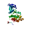

 Sample preparation
 / Beamline: 8.2.2 / Wavelength: 0.9537 Å
 Processing