- PDB-3kk7: Crystal structure of Putative cell invasion protein with MAC/Perf... -
+
Open data
ID or keywords:
Loading...
-
Basic information
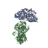
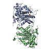
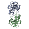
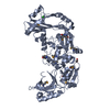
Entry
Database: PDB / ID: 3kk7
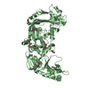
Title
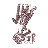
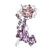
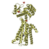
Crystal structure of Putative cell invasion protein with MAC/Perforin domain (NP_812351.1) from BACTERIODES THETAIOTAOMICRON VPI-5482 at 2.46 A resolution
Components
Putative cell invasion protein with MAC/Perforin domain
Keywords
CELL INVASION / Putative cell invasion protein with MAC/Perforin domain / Structural Genomics / Joint Center for Structural Genomics / JCSG / Protein Structure Initiative / PSI-2
Component-ID: 1 / Ens-ID: 1 / Beg auth comp-ID: THR / Beg label comp-ID: THR / End auth comp-ID: LEU / End label comp-ID: LEU / Refine code: 6 / Auth seq-ID: 36 - 558 / Label seq-ID: 19 - 541
Dom-ID
Auth asym-ID
Label asym-ID
1
A
A
2
B
B
Details
ANALYTICAL SIZE EXCLUSION CHROMATOGRAPHY WITH STATIC LIGHT SCATTERING AND CRYSTAL PACKING SUPPORT THE ASSIGNMENT OF A MONOMER AS THE SIGNIFICANT OLIGOMERIZATION STATE IN SOLUTION.
-
Components
#1: Protein
PutativecellinvasionproteinwithMAC/Perforindomain
Mass: 62007.379 Da / Num. of mol.: 2 Source method: isolated from a genetically manipulated source Source: (gene. exp.) Bacteroides thetaiotaomicron VPI-5482 (bacteria) Gene: BT_3439 / Plasmid: SpeedET / Production host: Escherichia Coli (E. coli) / Strain (production host): HK100 / References: UniProt: Q8A267
Mass: 18.015 Da / Num. of mol.: 239 / Source method: isolated from a natural source / Formula: H2O
Has protein modification
Y
Sequence details
THE CONSTRUCT (RESIDUES 19-558) WAS EXPRESSED WITH A PURIFICATION TAG MGSDKIHHHHHHENLYFQG. THE TAG ...THE CONSTRUCT (RESIDUES 19-558) WAS EXPRESSED WITH A PURIFICATION TAG MGSDKIHHHHHHENLYFQG. THE TAG WAS REMOVED WITH TEV PROTEASE LEAVING ONLY A GLYCINE (0) FOLLOWED BY THE TARGET SEQUENCE.
-
Experimental details
-
Experiment
Experiment
Method: X-RAY DIFFRACTION / Number of used crystals: 1
-
Sample preparation
Crystal
Density Matthews: 2.78 Å3/Da / Density % sol: 55.74 %
Crystal grow
Temperature: 277 K / Method: vapor diffusion, sitting drop / pH: 6.7 Details: 5.0000% 2-methyl-2,4-pentanediol, 12.0000% polyethylene glycol 6000, 0.1M HEPES pH 6.7, NANODROP', VAPOR DIFFUSION, SITTING DROP, temperature 277K
Resolution: 2.46→49.386 Å / Num. obs: 49779 / % possible obs: 97.3 % / Redundancy: 6.9 % / Biso Wilson estimate: 52.436 Å2 / Rmerge(I) obs: 0.123 / Rsym value: 0.123 / Net I/σ(I): 10.6
Reflection shell
Diffraction-ID: 1
Resolution (Å)
Redundancy (%)
Rmerge(I) obs
Mean I/σ(I) obs
Num. measured all
Num. unique all
Rsym value
% possible all
2.46-2.59
5.9
0.752
2.3
40706
6903
0.752
94.2
2.59-2.75
7.3
0.546
3.7
50621
6972
0.546
99.9
2.75-2.94
7.4
0.362
5.5
48478
6591
0.362
99.8
2.94-3.17
7.4
0.22
8.4
45109
6126
0.22
99.8
3.17-3.47
7.3
0.144
11.6
41087
5639
0.144
99.8
3.47-3.88
5.9
0.136
12.1
25745
4368
0.136
84.7
3.88-4.48
6.9
0.065
19.4
30873
4464
0.065
98.1
4.48-5.49
7.1
0.051
21.7
27703
3902
0.051
99.9
5.49-7.77
6.9
0.056
20.6
21239
3056
0.056
99.8
7.77-49.39
6.3
0.036
26.4
11003
1758
0.036
99
-
Phasing
Phasing
Method: MAD
-
Processing
Software
Name
Version
Classification
NB
REFMAC
5.5.0053
refinement
PHENIX
refinement
SHELX
phasing
MolProbity
3beta29
modelbuilding
SCALA
3.2.25
datascaling
PDB_EXTRACT
3.006
dataextraction
XDS
datareduction
SHELXD
phasing
autoSHARP
phasing
Refinement
Method to determine structure: MAD / Resolution: 2.46→49.386 Å / Cor.coef. Fo:Fc: 0.92 / Cor.coef. Fo:Fc free: 0.888 / Occupancy max: 1 / Occupancy min: 0.5 / SU B: 18.879 / SU ML: 0.19 / TLS residual ADP flag: LIKELY RESIDUAL / Cross valid method: THROUGHOUT / σ(F): 0 / ESU R: 0.405 / ESU R Free: 0.267 Stereochemistry target values: MAXIMUM LIKELIHOOD WITH PHASES Details: 1. HYDROGENS HAVE BEEN ADDED IN THE RIDING POSITIONS. 2. A MET-INHIBITION PROTOCOL WAS USED FOR SELENOMETHIONINE INCORPORATION DURING PROTEIN EXPRESSION. THE OCCUPANCY OF THE SE ATOMS IN THE ...Details: 1. HYDROGENS HAVE BEEN ADDED IN THE RIDING POSITIONS. 2. A MET-INHIBITION PROTOCOL WAS USED FOR SELENOMETHIONINE INCORPORATION DURING PROTEIN EXPRESSION. THE OCCUPANCY OF THE SE ATOMS IN THE MSE RESIDUES WAS REDUCED TO 0.75 FOR THE REDUCED SCATTERING POWER DUE TO PARTIAL S-MET INCORPORATION. 3. ATOM RECORDS CONTAIN RESIDUAL B FACTORS ONLY. 4. (4S)-2-METHY-2,4-PENTANEDIOL (MPD), ETHYLENE GLYCOL (EDO) AND CHLORIDE (CL) MODELED WERE PRESENT IN CRYSTLLIZATION/CRYO CONDITIONS.
Rfactor
Num. reflection
% reflection
Selection details
Rfree
0.252
2514
5.1 %
RANDOM
Rwork
0.209
-
-
-
obs
0.211
49764
97.25 %
-
Solvent computation
Ion probe radii: 0.8 Å / Shrinkage radii: 0.8 Å / VDW probe radii: 1.2 Å / Solvent model: MASK
In the structure databanks used in Yorodumi, some data are registered as the other names, "COVID-19 virus" and "2019-nCoV". Here are the details of the virus and the list of structure data.
Jan 31, 2019. EMDB accession codes are about to change! (news from PDBe EMDB page)
EMDB accession codes are about to change! (news from PDBe EMDB page)
The allocation of 4 digits for EMDB accession codes will soon come to an end. Whilst these codes will remain in use, new EMDB accession codes will include an additional digit and will expand incrementally as the available range of codes is exhausted. The current 4-digit format prefixed with “EMD-” (i.e. EMD-XXXX) will advance to a 5-digit format (i.e. EMD-XXXXX), and so on. It is currently estimated that the 4-digit codes will be depleted around Spring 2019, at which point the 5-digit format will come into force.
The EM Navigator/Yorodumi systems omit the EMD- prefix.
Related info.:Q: What is EMD? / ID/Accession-code notation in Yorodumi/EM Navigator
Yorodumi is a browser for structure data from EMDB, PDB, SASBDB, etc.
This page is also the successor to EM Navigator detail page, and also detail information page/front-end page for Omokage search.
The word "yorodu" (or yorozu) is an old Japanese word meaning "ten thousand". "mi" (miru) is to see.
Related info.:EMDB / PDB / SASBDB / Comparison of 3 databanks / Yorodumi Search / Aug 31, 2016. New EM Navigator & Yorodumi / Yorodumi Papers / Jmol/JSmol / Function and homology information / Changes in new EM Navigator and Yorodumi
 Movie
Movie Controller

 Yorodumi
 Open data
 Basic information
 Components
 Keywords
 Function and homology information
 Bacteroides thetaiotaomicron VPI-5482 (bacteria)
 X-RAY DIFFRACTION /
  Authors
 Citation
 Structure visualization
 Downloads & links
 Other downloads
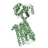


 PDBj
PDBj


 Assembly

 Mass: 35.453 Da / Num. of mol.: 1 / Source method: obtained synthetically / Formula: Cl
 Mass: 62.068 Da / Num. of mol.: 3 / Source method: obtained synthetically / Formula: C2H6O2
 Mass: 118.174 Da / Num. of mol.: 1 / Source method: obtained synthetically / Formula: C6H14O2 / Comment: precipitant*YM
 Mass: 18.015 Da / Num. of mol.: 239 / Source method: isolated from a natural source / Formula: H2O
 Sample preparation
 / Beamline: BL9-2 / Wavelength: 0.97908,0.91837,0.97922
 Processing