 Movie
Movie Controller

[English] 日本語
 Yorodumi
- PDB-3k9y: Crystal structure of rat mitochondrial P450 24A1 S57D in complex ... -
+ Open data

- Basic information
| Entry | Database: PDB / ID: 3k9y | ||||||
|---|---|---|---|---|---|---|---|
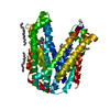
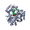
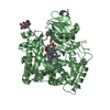
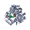
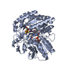
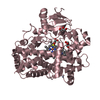
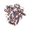
| Title | Crystal structure of rat mitochondrial P450 24A1 S57D in complex with CYMAL-5 | ||||||
 Components Components | 1,25-dihydroxyvitamin D(3) 24-hydroxylase, mitochondrial | ||||||
 Keywords Keywords | OXIDOREDUCTASE / MITOCHONDRIAL CYTOCHROME P450 / MONOTOPIC MEMBRANE PROTEIN / MONOOXYGENASE / VITAMIN D HORMONE METABOLISM / ADRENODOXIN | ||||||
| Function / homology |  Function and homology information Function and homology informationvitamin D3 24-hydroxylase / 25-hydroxycholecalciferol-24-hydroxylase activity / 1-alpha,25-dihydroxyvitamin D3 24-hydroxylase activity / 25-hydroxycholecalciferol-23-hydroxylase activity / Vitamins / Vitamin D (calciferol) metabolism / 1-alpha,25-dihydroxyvitamin D3 23-hydroxylase activity / vitamin D 25-hydroxylase activity / vitamin D 24-hydroxylase activity / vitamin D catabolic process ...vitamin D3 24-hydroxylase / 25-hydroxycholecalciferol-24-hydroxylase activity / 1-alpha,25-dihydroxyvitamin D3 24-hydroxylase activity / 25-hydroxycholecalciferol-23-hydroxylase activity / Vitamins / Vitamin D (calciferol) metabolism / 1-alpha,25-dihydroxyvitamin D3 23-hydroxylase activity / vitamin D 25-hydroxylase activity / vitamin D 24-hydroxylase activity / vitamin D catabolic process / vitamin D metabolic process / cellular response to vitamin D / response to vitamin D / osteoblast differentiation / iron ion binding / heme binding / mitochondrion Similarity search - Function | ||||||
| Biological species |  | ||||||
| Method |  X-RAY DIFFRACTION / SYNCHROTRON / MOLECULAR REPLACEMENT / Resolution: 2.8 Å X-RAY DIFFRACTION / SYNCHROTRON / MOLECULAR REPLACEMENT / Resolution: 2.8 Å | ||||||
 Authors Authors | Annalora, A.J. / Goodin, D.B. / Hong, W. / Zhang, Q. / Johnson, E.F. / Stout, C.D. | ||||||
 Citation Citation | Journal: J.Mol.Biol. / Year: 2010 Title: Crystal structure of CYP24A1, a mitochondrial cytochrome P450 involved in vitamin D metabolism. Authors: Annalora, A.J. / Goodin, D.B. / Hong, W.X. / Zhang, Q. / Johnson, E.F. / Stout, C.D. | ||||||
| History |
|
- Structure visualization
| Structure viewer | Molecule: MolmilJmol/JSmol |
|---|
- Downloads & links
-Download
| PDBx/mmCIF format | 3k9y.cif.gz | 188.1 KB | Display | PDBx/mmCIF format |
|---|---|---|---|---|
| PDB format | pdb3k9y.ent.gz | 150.4 KB | Display | PDB format |
| PDBx/mmJSON format | 3k9y.json.gz | Tree view | PDBx/mmJSON format | |
| Others |  Other downloads Other downloads |
-Validation report
| Arichive directory | https://data.pdbj.org/pub/pdb/validation_reports/k9/3k9yftp://data.pdbj.org/pub/pdb/validation_reports/k9/3k9y | HTTPS FTP |
|---|
-Related structure data
| Related structure data |  3k9vC  1tqnS C: citing same article ( S: Starting model for refinement |
|---|---|
| Similar structure data |


-Links
 PDBj
PDBj- Assembly
| Deposited unit | 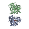
| ||||||||
|---|---|---|---|---|---|---|---|---|---|
| 1 | 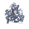
| ||||||||
| 2 | 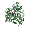
| ||||||||
| 3 | 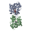
| ||||||||
| Unit cell |
|
-Components
| #1: Protein | Mass: 55933.773 Da / Num. of mol.: 2 / Fragment: residues 34-514 / Mutation: S57D Source method: isolated from a genetically manipulated source Source: (gene. exp.)  References: UniProt: Q09128, Oxidoreductases; Acting on paired donors, with incorporation or reduction of molecular oxygen; With NADH or NADPH as one donor, and incorporation of one atom of oxygen into the other donor #2: Chemical |   Mass: 616.487 Da / Num. of mol.: 2 / Source method: obtained synthetically / Formula: C34H32FeN4O4 Mass: 616.487 Da / Num. of mol.: 2 / Source method: obtained synthetically / Formula: C34H32FeN4O4#3: Chemical |   Mass: 494.573 Da / Num. of mol.: 2 / Source method: obtained synthetically / Formula: C23H42O11 / Comment: detergent*YM Mass: 494.573 Da / Num. of mol.: 2 / Source method: obtained synthetically / Formula: C23H42O11 / Comment: detergent*YM |
|---|
-Experimental details
-Experiment
| Experiment | Method: X-RAY DIFFRACTION / Number of used crystals: 1 |
|---|
- Sample preparation
| Crystal | Density Matthews: 3.01 Å3/Da / Density % sol: 59.09 % |
|---|---|
| Crystal grow | Temperature: 290 K / Method: vapor diffusion, sitting drop / pH: 8.5 Details: 15% PEG 3350, 0.1 M Tris-HCl, 0.2M potassium thiocyanate, 0.05M CaCl, pH 8.5, VAPOR DIFFUSION, SITTING DROP, temperature 290K |
-Data collection
| Diffraction source | Source: SYNCHROTRON / Site: SSRL  / Beamline: BL9-2 / Wavelength: 0.97946 Å / Beamline: BL9-2 / Wavelength: 0.97946 Å |
|---|---|
| Detector | Type: MARMOSAIC 325 mm CCD / Detector: CCD / Date: Nov 7, 2007 |
| Radiation | Protocol: SINGLE WAVELENGTH / Monochromatic (M) / Laue (L): M / Scattering type: x-ray |
| Radiation wavelength | Wavelength: 0.97946 Å / Relative weight: 1 |
| Reflection | Resolution: 2.8→91.67 Å / Num. all: 32727 / % possible obs: 97 % / Redundancy: 2.5 % / Biso Wilson estimate: 67.645 Å2 / Rmerge(I) obs: 0.166 / Rsym value: 0.166 / Net I/σ(I): 7.9 |
| Reflection shell | Resolution: 2.8→2.87 Å / Redundancy: 2.5 % / Rmerge(I) obs: 1.332 / Mean I/σ(I) obs: 1.1 / Num. unique all: 1768 / Rsym value: 1.642 / % possible all: 70.6 |
- Processing
| Software |
| ||||||||||||||||||||
|---|---|---|---|---|---|---|---|---|---|---|---|---|---|---|---|---|---|---|---|---|---|
| Refinement | Method to determine structure: MOLECULAR REPLACEMENT Starting model: 1TQN Resolution: 2.8→50 Å
| ||||||||||||||||||||
| Displacement parameters | Biso mean: 77.151 Å2
| ||||||||||||||||||||
| Refinement step | Cycle: LAST / Resolution: 2.8→50 Å
| ||||||||||||||||||||
| Refine LS restraints |
|
