 Movie
Movie Controller

[English] 日本語
 Yorodumi
- PDB-3k81: Structure of the central interaction protein from the Trypanosoma... -
+ Open data

- Basic information
| Entry | Database: PDB / ID: 3k81 | ||||||
|---|---|---|---|---|---|---|---|
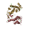
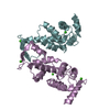
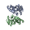
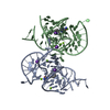
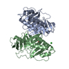
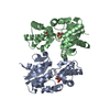
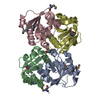
| Title | Structure of the central interaction protein from the Trypanosoma brucei editosome in complex with single domain antibodies | ||||||
 Components Components |
| ||||||
 Keywords Keywords | Immune System / RNA Binding Protein / KREPA6 / VHH / Single domain antibody | ||||||
| Function / homology |  Function and homology information Function and homology informationRNA modification / mitochondrial mRNA editing complex / kinetoplast / single-stranded DNA binding / endonuclease activity / RNA binding / nucleus Similarity search - Function | ||||||
| Biological species |   | ||||||
| Method |  X-RAY DIFFRACTION / SYNCHROTRON / MOLECULAR REPLACEMENT / Resolution: 3.4 Å X-RAY DIFFRACTION / SYNCHROTRON / MOLECULAR REPLACEMENT / Resolution: 3.4 Å | ||||||
 Authors Authors | Park, Y.-J. / Hol, W. | ||||||
 Citation Citation | Journal: J.Struct.Biol. / Year: 2011 Title: Structures of a key interaction protein from the Trypanosoma brucei editosome in complex with single domain antibodies. Authors: Wu, M. / Park, Y.J. / Pardon, E. / Turley, S. / Hayhurst, A. / Deng, J. / Steyaert, J. / Hol, W.G. | ||||||
| History |
|
- Structure visualization
| Structure viewer | Molecule: MolmilJmol/JSmol |
|---|
- Downloads & links
-Download
| PDBx/mmCIF format | 3k81.cif.gz | 178.6 KB | Display | PDBx/mmCIF format |
|---|---|---|---|---|
| PDB format | pdb3k81.ent.gz | 143.1 KB | Display | PDB format |
| PDBx/mmJSON format | 3k81.json.gz | Tree view | PDBx/mmJSON format | |
| Others |  Other downloads Other downloads |
-Validation report
| Arichive directory | https://data.pdbj.org/pub/pdb/validation_reports/k8/3k81ftp://data.pdbj.org/pub/pdb/validation_reports/k8/3k81 | HTTPS FTP |
|---|
-Related structure data
| Related structure data | 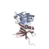 3k7uSC 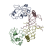 3k80C S: Starting model for refinement C: citing same article ( |
|---|---|
| Similar structure data |

-Links
 PDBj
PDBj

- Assembly
| Deposited unit | 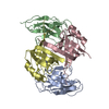
| ||||||||
|---|---|---|---|---|---|---|---|---|---|
| 1 |
| ||||||||
| Unit cell |
|
-Components
| #1: Protein | Mass: 18113.566 Da / Num. of mol.: 2 / Fragment: KREPA6 Source method: isolated from a genetically manipulated source Source: (gene. exp.)  #2: Antibody | Mass: 13967.477 Da / Num. of mol.: 2 / Fragment: single domain antibody VHH Source method: isolated from a genetically manipulated source Source: (gene. exp.) Has protein modification | Y | |
|---|
-Experimental details
-Experiment
| Experiment | Method: X-RAY DIFFRACTION / Number of used crystals: 1 |
|---|
- Sample preparation
| Crystal | Density Matthews: 1.95 Å3/Da / Density % sol: 36.97 % |
|---|---|
| Crystal grow | Temperature: 293 K / Method: vapor diffusion, sitting drop / pH: 4.2 Details: 15% PEG3350, 0.1M Citrate pH.4.2, VAPOR DIFFUSION, SITTING DROP, temperature 293K |
-Data collection
| Diffraction | Mean temperature: 77 K |
|---|---|
| Diffraction source | Source: SYNCHROTRON / Site: SSRL  / Beamline: BL9-2 / Wavelength: 0.9795 Å / Beamline: BL9-2 / Wavelength: 0.9795 Å |
| Detector | Type: MARMOSAIC 325 mm CCD / Detector: CCD / Date: Apr 1, 2009 Details: Flat collimating mirror, double crystal monochromator, toroid focusing mirror |
| Radiation | Monochromator: Double crystal monochromator / Protocol: SINGLE WAVELENGTH / Monochromatic (M) / Laue (L): M / Scattering type: x-ray |
| Radiation wavelength | Wavelength: 0.9795 Å / Relative weight: 1 |
| Reflection | Resolution: 3.4→50 Å / Num. obs: 6402 / % possible obs: 92.3 % / Observed criterion σ(I): 3 / Redundancy: 3.3 % / Biso Wilson estimate: 64.9 Å2 / Rmerge(I) obs: 0.112 / Rsym value: 0.112 / Net I/σ(I): 8.6 |
| Reflection shell | Resolution: 3.4→3.52 Å / Redundancy: 2.3 % / Rmerge(I) obs: 0.338 / Mean I/σ(I) obs: 2 / Num. unique all: 423 / Rsym value: 0.338 / % possible all: 61.2 |
- Processing
| Software |
| ||||||||||||||||||||||||||||||||||||||||
|---|---|---|---|---|---|---|---|---|---|---|---|---|---|---|---|---|---|---|---|---|---|---|---|---|---|---|---|---|---|---|---|---|---|---|---|---|---|---|---|---|---|
| Refinement | Method to determine structure: MOLECULAR REPLACEMENT Starting model: PDB ENTRY 3K7U Resolution: 3.4→33.467 Å / SU ML: 0.73 / σ(F): 1.38 / Stereochemistry target values: ML
| ||||||||||||||||||||||||||||||||||||||||
| Solvent computation | Shrinkage radii: 0.9 Å / VDW probe radii: 1.11 Å / Solvent model: FLAT BULK SOLVENT MODEL / Bsol: 114.052 Å2 / ksol: 0.346 e/Å3 | ||||||||||||||||||||||||||||||||||||||||
| Refinement step | Cycle: LAST / Resolution: 3.4→33.467 Å
| ||||||||||||||||||||||||||||||||||||||||
| Refine LS restraints |
| ||||||||||||||||||||||||||||||||||||||||
| LS refinement shell |
| ||||||||||||||||||||||||||||||||||||||||
| Refinement TLS params. | Method: refined / Origin x: 24.3208 Å / Origin y: 11.4461 Å / Origin z: -23.0129 Å
| ||||||||||||||||||||||||||||||||||||||||
| Refinement TLS group | Selection details: all |
