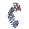
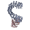
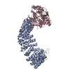
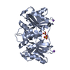
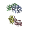
[English] 日本語
Yorodumi
- PDB-3k7w: Protein phosphatase 2A core complex bound to dinophysistoxin-2 -

+
Open data


ID or keywords:

Loading...

-
Basic information

Entry
Database: PDB / ID: 3k7w
TitleProtein phosphatase 2A core complex bound to dinophysistoxin-2
Components(Serine/threonine-protein phosphatase 2A ...) x 2
KeywordsSIGNALING PROTEIN/HYDROLASE / PROTEIN-PROTEIN COMPLEX / HEAT REPEAT / SIGNALING PROTEIN / PHOSPHATASE / LIGAND SPECIFICITY / Acetylation / Polymorphism / Centromere / Cytoplasm / Cytoskeleton / Hydrolase / Iron / Manganese / Metal-binding / Methylation / Nucleus / Phosphoprotein / Protein phosphatase / SIGNALING PROTEIN-HYDROLASE complex
Function / homology
Function and homology information


meiotic spindle elongation / Integration of energy metabolism / PP2A-mediated dephosphorylation of key metabolic factors / regulation of microtubule binding / MASTL Facilitates Mitotic Progression / mitotic sister chromatid separation / regulation of meiotic cell cycle process involved in oocyte maturation / protein phosphatase type 2A complex / meiotic sister chromatid cohesion, centromeric / : ...meiotic spindle elongation / Integration of energy metabolism / PP2A-mediated dephosphorylation of key metabolic factors / regulation of microtubule binding / MASTL Facilitates Mitotic Progression / mitotic sister chromatid separation / regulation of meiotic cell cycle process involved in oocyte maturation / protein phosphatase type 2A complex / meiotic sister chromatid cohesion, centromeric / : / positive regulation of microtubule binding / negative regulation of tyrosine phosphorylation of STAT protein / Regulation of glycolysis by fructose 2,6-bisphosphate metabolism / Inhibition of replication initiation of damaged DNA by RB1/E2F1 / female meiotic nuclear division / protein antigen binding / protein phosphatase regulator activity / ceramide metabolic process / peptidyl-threonine dephosphorylation / GABA receptor binding / negative regulation of epithelial to mesenchymal transition / peptidyl-serine dephosphorylation / APC truncation mutants have impaired AXIN binding / AXIN missense mutants destabilize the destruction complex / Truncations of AMER1 destabilize the destruction complex / Initiation of Nuclear Envelope (NE) Reformation / ERKs are inactivated / positive regulation of extrinsic apoptotic signaling pathway in absence of ligand / Beta-catenin phosphorylation cascade / Signaling by GSK3beta mutants / CTNNB1 S33 mutants aren't phosphorylated / CTNNB1 S37 mutants aren't phosphorylated / CTNNB1 S45 mutants aren't phosphorylated / CTNNB1 T41 mutants aren't phosphorylated / regulation of Wnt signaling pathway / Disassembly of the destruction complex and recruitment of AXIN to the membrane / regulation of growth / negative regulation of glycolytic process through fructose-6-phosphate / positive regulation of NLRP3 inflammasome complex assembly / myosin phosphatase activity / protein serine/threonine phosphatase activity / CTLA4 inhibitory signaling / Platelet sensitization by LDL / negative regulation of MAPK cascade / protein-serine/threonine phosphatase / regulation of cell differentiation / T cell homeostasis / ERK/MAPK targets / regulation of G1/S transition of mitotic cell cycle / regulation of DNA replication / phosphoprotein phosphatase activity / mesoderm development / chromosome, centromeric region / DARPP-32 events / lateral plasma membrane / Nonsense Mediated Decay (NMD) enhanced by the Exon Junction Complex (EJC) / Amplification of signal from unattached kinetochores via a MAD2 inhibitory signal / regulation of cell adhesion / negative regulation of phosphatidylinositol 3-kinase/protein kinase B signal transduction / Cyclin A/B1/B2 associated events during G2/M transition / Mitotic Prometaphase / EML4 and NUDC in mitotic spindle formation / Loss of Nlp from mitotic centrosomes / Loss of proteins required for interphase microtubule organization from the centrosome / Recruitment of mitotic centrosome proteins and complexes / Resolution of Sister Chromatid Cohesion / Recruitment of NuMA to mitotic centrosomes / Anchoring of the basal body to the plasma membrane / AURKA Activation by TPX2 / protein dephosphorylation / response to organic substance / RNA splicing / meiotic cell cycle / protein tyrosine phosphatase activity / chromosome segregation / RHO GTPases Activate Formins / response to lead ion / regulation of protein phosphorylation / Spry regulation of FGF signaling / RAF activation / tau protein binding / Degradation of beta-catenin by the destruction complex / PKR-mediated signaling / positive regulation of protein serine/threonine kinase activity / spindle pole / negative regulation of cell growth / Negative regulation of MAPK pathway / Separation of Sister Chromatids / microtubule cytoskeleton / Cyclin D associated events in G1 / Regulation of PLK1 Activity at G2/M Transition / Regulation of TP53 Degradation / mitotic cell cycle / PI5P, PP2A and IER3 Regulate PI3K/AKT Signaling / protein-containing complex assembly / neuron projection / membrane raft / protein heterodimerization activity / neuronal cell body / dendrite
Similarity search - Function
: / HEAT repeat / HEAT repeat / Serine/threonine specific protein phosphatases signature. / Protein phosphatase 2A homologues, catalytic domain. / Serine/threonine-specific protein phosphatase/bis(5-nucleosyl)-tetraphosphatase / HEAT repeat profile. / HEAT, type 2 / HEAT repeats / Metallo-dependent phosphatases ...: / HEAT repeat / HEAT repeat / Serine/threonine specific protein phosphatases signature. / Protein phosphatase 2A homologues, catalytic domain. / Serine/threonine-specific protein phosphatase/bis(5-nucleosyl)-tetraphosphatase / HEAT repeat profile. / HEAT, type 2 / HEAT repeats / Metallo-dependent phosphatases / Purple Acid Phosphatase; chain A, domain 2 / Calcineurin-like phosphoesterase domain, ApaH type / Calcineurin-like phosphoesterase / Leucine-rich Repeat Variant / Leucine-rich Repeat Variant / Metallo-dependent phosphatase-like / Armadillo-like helical / 4-Layer Sandwich / Alpha Horseshoe / Armadillo-type fold / Mainly Alpha / Alpha Beta
Similarity search - Domain/homology
: / Chem-XT2 / Serine/threonine-protein phosphatase 2A 65 kDa regulatory subunit A alpha isoform / Serine/threonine-protein phosphatase 2A catalytic subunit alpha isoform
Similarity search - Component
Biological speciesHomo sapiens (human)
MethodX-RAY DIFFRACTION / SYNCHROTRON / Rigid body refinement / Resolution: 2.96 Å
AuthorsJeffrey, P.D. / Huhn, J. / Shi, Y.
CitationJournal: Chem.Res.Toxicol. / Year: 2009
Title: A structural basis for the reduced toxicity of dinophysistoxin-2.
Authors: Huhn, J. / Jeffrey, P.D. / Larsen, K. / Rundberget, T. / Rise, F. / Cox, N.R. / Arcus, V. / Shi, Y. / Miles, C.O.
History
DepositionOct 13, 2009Deposition site: RCSB / Processing site: RCSB
Revision 1.0Nov 3, 2009Provider: repository / Type: Initial release
Revision 1.1Jul 13, 2011Group: Version format compliance
Revision 1.2Sep 6, 2023Group: Data collection / Database references ...Data collection / Database references / Derived calculations / Refinement description / Structure summary
Category: chem_comp / chem_comp_atom ...chem_comp / chem_comp_atom / chem_comp_bond / database_2 / entity / pdbx_entity_nonpoly / pdbx_initial_refinement_model / pdbx_struct_conn_angle / struct_conn / struct_site
Item: _chem_comp.name / _database_2.pdbx_DOI ..._chem_comp.name / _database_2.pdbx_DOI / _database_2.pdbx_database_accession / _entity.pdbx_description / _pdbx_entity_nonpoly.name / _pdbx_struct_conn_angle.ptnr1_auth_comp_id / _pdbx_struct_conn_angle.ptnr1_auth_seq_id / _pdbx_struct_conn_angle.ptnr1_label_atom_id / _pdbx_struct_conn_angle.ptnr1_label_comp_id / _pdbx_struct_conn_angle.ptnr1_label_seq_id / _pdbx_struct_conn_angle.ptnr2_auth_seq_id / _pdbx_struct_conn_angle.ptnr2_label_asym_id / _pdbx_struct_conn_angle.ptnr3_auth_comp_id / _pdbx_struct_conn_angle.ptnr3_auth_seq_id / _pdbx_struct_conn_angle.ptnr3_label_atom_id / _pdbx_struct_conn_angle.ptnr3_label_comp_id / _pdbx_struct_conn_angle.ptnr3_label_seq_id / _pdbx_struct_conn_angle.value / _struct_conn.pdbx_dist_value / _struct_conn.ptnr1_auth_comp_id / _struct_conn.ptnr1_auth_seq_id / _struct_conn.ptnr1_label_atom_id / _struct_conn.ptnr1_label_comp_id / _struct_conn.ptnr1_label_seq_id / _struct_conn.ptnr2_auth_seq_id / _struct_conn.ptnr2_label_asym_id / _struct_site.pdbx_auth_asym_id / _struct_site.pdbx_auth_comp_id / _struct_site.pdbx_auth_seq_id

-
Structure visualization

Structure viewerMolecule:
MolmilJmol/JSmol

Downloads & links

-
Assembly

Deposited unit
A: Serine/threonine-protein phosphatase 2A 65 kDa regulatory subunit A alpha isoform
C: Serine/threonine-protein phosphatase 2A catalytic subunit alpha isoform
hetero molecules


Theoretical massNumber of molelcules
Total (without water)102,2188
Polymers101,0142
Non-polymers1,2036
Water45025
1


  • Idetical with deposited unit
  • defined by author&software
TypeNameSymmetry operationNumber
identity operation1_555x,y,z1
Buried area2720 Å2
ΔGint-59 kcal/mol
Surface area35980 Å2
MethodPISA
Unit cell
Length a, b, c (Å)93.410, 197.240, 201.650
Angle α, β, γ (deg.)90.00, 90.00, 90.00
Int Tables number23
Space group name H-MI222

-
Components

-
Serine/threonine-protein phosphatase 2A ... , 2 types, 2 molecules AC

#1: Protein Serine/threonine-protein phosphatase 2A 65 kDa regulatory subunit A alpha isoform / PP2A / subunit A / PR65-alpha isoform / PP2A / subunit A / R1-alpha isoform / Medium tumor antigen- ...PP2A / subunit A / PR65-alpha isoform / PP2A / subunit A / R1-alpha isoform / Medium tumor antigen-associated 61 kDa protein


Mass: 65378.344 Da / Num. of mol.: 1
Source method: isolated from a genetically manipulated source
Source: (gene. exp.) Homo sapiens (human) / Gene: PPP2R1A / Plasmid: PGEX-2T / Production host: Escherichia coli (E. coli) / Strain (production host): BL21(DE3) / References: UniProt: P30153
#2: Protein Serine/threonine-protein phosphatase 2A catalytic subunit alpha isoform / PP2A-alpha / Replication protein C / RP-C


Mass: 35636.152 Da / Num. of mol.: 1
Source method: isolated from a genetically manipulated source
Source: (gene. exp.) Homo sapiens (human) / Gene: PPP2CA / Production host: 
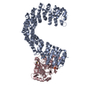
Spodoptera frugiperda (fall armyworm) / Strain (production host): HI-5
References: UniProt: P67775, protein-serine/threonine phosphatase

-
Non-polymers , 4 types, 31 molecules

#3: Chemical ChemComp-SO4 / SULFATE ION / Sulfate


Mass: 96.063 Da / Num. of mol.: 3 / Source method: obtained synthetically / Formula: SO4
#4: Chemical ChemComp-XT2 / (2R)-2-hydroxy-3-[(2S,5R,6R,8S)-5-hydroxy-8-{(1R,2E)-3-[(2R,4a'R,5R,6'S,8'R,8a'S)-8'-hydroxy-6'-{(1S,3S)-1-hydroxy-3-[( 2S,6R,11S)-11-methyl-1,7-dioxaspiro[5.5]undec-2-yl]butyl}-7'-methylideneoctahydro-3H,3'H-spiro[furan-2,2'-pyrano[3,2-b]p yran]-5-yl]-1-methylprop-2-en-1-yl}-10-methyl-1,7-dioxaspiro[5.5]undec-10-en-2-yl]-2-methylpropanoic acid / DINOPHYSISTOXIN-2


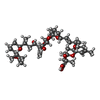
Mass: 805.003 Da / Num. of mol.: 1 / Source method: obtained synthetically / Formula: C44H68O13
#5: Chemical ChemComp-MN / MANGANESE (II) ION


Mass: 54.938 Da / Num. of mol.: 2 / Source method: obtained synthetically / Formula: Mn
#6: Water ChemComp-HOH / water / Water


Mass: 18.015 Da / Num. of mol.: 25 / Source method: isolated from a natural source / Formula: H2O

-
Experimental details

-
Experiment

ExperimentMethod: X-RAY DIFFRACTION / Number of used crystals: 1

-
Sample preparation

CrystalDensity Matthews: 4.6 Å3/Da / Density % sol: 73.25 %
Crystal growTemperature: 293 K / Method: vapor diffusion, hanging drop / pH: 8.5
Details: 0.2 M LITHIUM SULFATE, 1.5 M AMMONIUM SULFATE, 0.1 M TRIS-CL, pH 8.5, VAPOR DIFFUSION, HANGING DROP, temperature 293K

-
Data collection

DiffractionMean temperature: 100 K
Diffraction sourceSource: SYNCHROTRON / Site: NSLS / Beamline: X29A / Wavelength: 1.0809 Å
DetectorType: ADSC QUANTUM 315 / Detector: CCD / Date: Aug 2, 2007
RadiationMonochromator: Si(111) / Protocol: SINGLE WAVELENGTH / Monochromatic (M) / Laue (L): M / Scattering type: x-ray
Radiation wavelengthWavelength: 1.0809 Å / Relative weight: 1
ReflectionResolution: 2.95→50 Å / Num. all: 39083 / Num. obs: 39083 / % possible obs: 99.8 % / Observed criterion σ(I): -3 / Redundancy: 3.9 % / Biso Wilson estimate: 139.4 Å2 / Rsym value: 0.082
Reflection shellResolution: 2.95→3.06 Å / Redundancy: 4 % / Rsym value: 0.439 / % possible all: 99.9

-
Processing

Software
NameClassification
CBASSdata collection
CNSrefinement
DENZOdata reduction
SCALEPACKdata scaling
CNSphasing
RefinementMethod to determine structure: Rigid body refinement
Starting model: pdb entry 2IE4
Resolution: 2.96→29.81 Å / Rfactor Rfree error: 0.006 / Data cutoff high absF: 2439426.18 / Data cutoff low absF: 0 / Isotropic thermal model: RESTRAINED / Cross valid method: THROUGHOUT / σ(F): 0 / Stereochemistry target values: Engh & Huber / Details: BULK SOLVENT MODEL USED
RfactorNum. reflection% reflectionSelection details
Rfree0.263 1918 4.9 %RANDOM
Rwork0.222 ---
obs0.222 39046 99.5 %-
all-39046 --
Solvent computationSolvent model: FLAT MODEL / Bsol: 24.8995 Å2 / ksol: 0.3 e/Å3
Displacement parametersBiso mean: 65.8 Å2
Baniso -1Baniso -2Baniso -3
1--6.17 Å20 Å20 Å2
2---4.76 Å20 Å2
3---10.93 Å2
Refine analyze
FreeObs
Luzzati coordinate error0.48 Å0.39 Å
Luzzati d res low-5 Å
Luzzati sigma a0.71 Å0.57 Å
Refinement stepCycle: LAST / Resolution: 2.96→29.81 Å
ProteinNucleic acidLigandSolventTotal
Num. atoms6849 0 74 25 6948
Refine LS restraints
Refine-IDTypeDev ideal
X-RAY DIFFRACTIONc_bond_d0.009
X-RAY DIFFRACTIONc_bond_d_na
X-RAY DIFFRACTIONc_bond_d_prot
X-RAY DIFFRACTIONc_angle_d
X-RAY DIFFRACTIONc_angle_d_na
X-RAY DIFFRACTIONc_angle_d_prot
X-RAY DIFFRACTIONc_angle_deg1.5
X-RAY DIFFRACTIONc_angle_deg_na
X-RAY DIFFRACTIONc_angle_deg_prot
X-RAY DIFFRACTIONc_dihedral_angle_d20.7
X-RAY DIFFRACTIONc_dihedral_angle_d_na
X-RAY DIFFRACTIONc_dihedral_angle_d_prot
X-RAY DIFFRACTIONc_improper_angle_d0.98
X-RAY DIFFRACTIONc_improper_angle_d_na
X-RAY DIFFRACTIONc_improper_angle_d_prot
X-RAY DIFFRACTIONc_mcbond_it
X-RAY DIFFRACTIONc_mcangle_it
X-RAY DIFFRACTIONc_scbond_it
X-RAY DIFFRACTIONc_scangle_it
LS refinement shellResolution: 2.96→3.13 Å / Rfactor Rfree error: 0.023 / Total num. of bins used: 6
RfactorNum. reflection% reflection
Rfree0.396 304 5 %
Rwork0.336 5721 -
obs--92.4 %
Xplor file
Refine-IDSerial noParam fileTopol file
X-RAY DIFFRACTION1protein_rep.paramprotein.top
X-RAY DIFFRACTION2dtx2.paramdtx2.top
X-RAY DIFFRACTION3water_rep.paramwater.top
X-RAY DIFFRACTION4ion.paramion.top
X-RAY DIFFRACTION5param_mn.inptopo_mn.inp

+
About Yorodumi

-
News

-
Feb 9, 2022. New format data for meta-information of EMDB entries

New format data for meta-information of EMDB entries

  • Version 3 of the EMDB header file is now the official format.
  • The previous official version 1.9 will be removed from the archive.

Related info.:EMDB header

External links:wwPDB to switch to version 3 of the EMDB data model

-
Aug 12, 2020. Covid-19 info

Covid-19 info

URL: https://pdbj.org/emnavi/covid19.php

New page: Covid-19 featured information page in EM Navigator.

Related info.:Covid-19 info / Mar 5, 2020. Novel coronavirus structure data

+
Mar 5, 2020. Novel coronavirus structure data

Novel coronavirus structure data

Related info.:Yorodumi Speices / Aug 12, 2020. Covid-19 info

External links:COVID-19 featured content - PDBj / Molecule of the Month (242):Coronavirus Proteases

+
Jan 31, 2019. EMDB accession codes are about to change! (news from PDBe EMDB page)

EMDB accession codes are about to change! (news from PDBe EMDB page)

  • The allocation of 4 digits for EMDB accession codes will soon come to an end. Whilst these codes will remain in use, new EMDB accession codes will include an additional digit and will expand incrementally as the available range of codes is exhausted. The current 4-digit format prefixed with “EMD-” (i.e. EMD-XXXX) will advance to a 5-digit format (i.e. EMD-XXXXX), and so on. It is currently estimated that the 4-digit codes will be depleted around Spring 2019, at which point the 5-digit format will come into force.
  • The EM Navigator/Yorodumi systems omit the EMD- prefix.

Related info.:Q: What is EMD? / ID/Accession-code notation in Yorodumi/EM Navigator

External links:EMDB Accession Codes are Changing Soon! / Contact to PDBj

+
Jul 12, 2017. Major update of PDB

Major update of PDB

  • wwPDB released updated PDB data conforming to the new PDBx/mmCIF dictionary.
  • This is a major update changing the version number from 4 to 5, and with Remediation, in which all the entries are updated.
  • In this update, many items about electron microscopy experimental information are reorganized (e.g. em_software).
  • Now, EM Navigator and Yorodumi are based on the updated data.

External links:wwPDB Remediation / Enriched Model Files Conforming to OneDep Data Standards Now Available in the PDB FTP Archive

-
Yorodumi

Thousand views of thousand structures

  • Yorodumi is a browser for structure data from EMDB, PDB, SASBDB, etc.
  • This page is also the successor to EM Navigator detail page, and also detail information page/front-end page for Omokage search.
  • The word "yorodu" (or yorozu) is an old Japanese word meaning "ten thousand". "mi" (miru) is to see.

Related info.:EMDB / PDB / SASBDB / Comparison of 3 databanks / Yorodumi Search / Aug 31, 2016. New EM Navigator & Yorodumi / Yorodumi Papers / Jmol/JSmol / Function and homology information / Changes in new EM Navigator and Yorodumi

Read more