 Movie
Movie Controller

[English] 日本語
 Yorodumi
- PDB-3k52: Crystal Structure of Isopentenyl Phosphate Kinase from M. jannasc... -
+ Open data

- Basic information
| Entry | Database: PDB / ID: 3k52 | ||||||
|---|---|---|---|---|---|---|---|
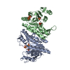
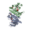
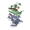
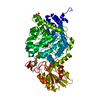
| Title | Crystal Structure of Isopentenyl Phosphate Kinase from M. jannaschii in complex with IP | ||||||
 Components Components | isopentenyl phosphate kinase | ||||||
 Keywords Keywords | TRANSFERASE / small molecule kinase / ATP-binding / Methanocaldococcus jannaschii / isopentenyl monophosphate / isopentenyl diphosphate / isoprenoid biosynthesis / mevalonate pathway / archaea | ||||||
| Function / homology |  Function and homology information Function and homology informationisopentenyl phosphate kinase / isopentenyl phosphate kinase activity / terpenoid biosynthetic process / kinase activity / ATP binding / cytosol Similarity search - Function | ||||||
| Biological species |   Methanocaldococcus jannaschii (archaea) Methanocaldococcus jannaschii (archaea) | ||||||
| Method |  X-RAY DIFFRACTION / SYNCHROTRON / MOLECULAR REPLACEMENT / Resolution: 2.7 Å X-RAY DIFFRACTION / SYNCHROTRON / MOLECULAR REPLACEMENT / Resolution: 2.7 Å | ||||||
| Model details | Crystal Structure of Isopentenyl Phosphate Kinase from M. jannaschii in complex with IP | ||||||
 Authors Authors | Dellas, N. / Noel, J.P. | ||||||
 Citation Citation | Journal: Acs Chem.Biol. / Year: 2010 Title: Mutation of archaeal isopentenyl phosphate kinase highlights mechanism and guides phosphorylation of additional isoprenoid monophosphates. Authors: Dellas, N. / Noel, J.P. | ||||||
| History |
|
- Structure visualization
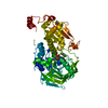
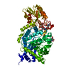
| Structure viewer | Molecule: MolmilJmol/JSmol |
|---|
- Downloads & links
-Download
| PDBx/mmCIF format | 3k52.cif.gz | 112.2 KB | Display | PDBx/mmCIF format |
|---|---|---|---|---|
| PDB format | pdb3k52.ent.gz | 87.9 KB | Display | PDB format |
| PDBx/mmJSON format | 3k52.json.gz | Tree view | PDBx/mmJSON format | |
| Others |  Other downloads Other downloads |
-Validation report
| Arichive directory | https://data.pdbj.org/pub/pdb/validation_reports/k5/3k52ftp://data.pdbj.org/pub/pdb/validation_reports/k5/3k52 | HTTPS FTP |
|---|
-Related structure data


-Links
 PDBj
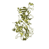
PDBj- Assembly
| Deposited unit | 
| ||||||||
|---|---|---|---|---|---|---|---|---|---|
| 1 | 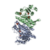
| ||||||||
| 2 | 
| ||||||||
| Unit cell |
|
-Components
| #1: Protein | Mass: 30009.746 Da / Num. of mol.: 2 Source method: isolated from a genetically manipulated source Source: (gene. exp.) Methanocaldococcus jannaschii (archaea)Gene: MJ0044 / Plasmid: pHIS8 / Production host:  #2: Chemical | 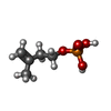  Mass: 166.112 Da / Num. of mol.: 2 / Source method: obtained synthetically / Formula: C5H11O4P Mass: 166.112 Da / Num. of mol.: 2 / Source method: obtained synthetically / Formula: C5H11O4P#3: Chemical | ChemComp-SO4 /   Mass: 96.063 Da / Num. of mol.: 4 / Source method: obtained synthetically / Formula: SO4 Mass: 96.063 Da / Num. of mol.: 4 / Source method: obtained synthetically / Formula: SO4#4: Water | ChemComp-HOH / |  Mass: 18.015 Da / Num. of mol.: 61 / Source method: isolated from a natural source / Formula: H2O Mass: 18.015 Da / Num. of mol.: 61 / Source method: isolated from a natural source / Formula: H2O |
|---|
-Experimental details
-Experiment
| Experiment | Method: X-RAY DIFFRACTION / Number of used crystals: 1 |
|---|
- Sample preparation
| Crystal | Density Matthews: 2.85 Å3/Da / Density % sol: 56.91 % |
|---|---|
| Crystal grow | Temperature: 298 K / Method: vapor diffusion, hanging drop Details: crystals were grown in 1.6M ammonium sulfate, transferred to 1.6M ammonium sulfate, 2mM IP, vapor diffusion, hanging drop, temperature 298K, VAPOR DIFFUSION, HANGING DROP |
-Data collection
| Diffraction | Mean temperature: 100 K |
|---|---|
| Diffraction source | Source: SYNCHROTRON / Site: ALS  / Beamline: 8.2.2 / Wavelength: 1 Å / Beamline: 8.2.2 / Wavelength: 1 Å |
| Detector | Type: ADSC QUANTUM 315 / Detector: CCD / Date: Aug 27, 2008 |
| Radiation | Monochromator: double crystal Si(111) / Protocol: SINGLE WAVELENGTH / Monochromatic (M) / Laue (L): M / Scattering type: x-ray |
| Radiation wavelength | Wavelength: 1 Å / Relative weight: 1 |
| Reflection | Resolution: 2.7→50 Å / Num. all: 19488 / Num. obs: 19309 / % possible obs: 99.1 % / Observed criterion σ(I): -3 / Redundancy: 5.88 % / Biso Wilson estimate: 54.186 Å2 / Rmerge(I) obs: 0.074 / Net I/σ(I): 16.53 |
| Reflection shell | Resolution: 2.7→2.8 Å / Redundancy: 5.81 % / Rmerge(I) obs: 0.526 / Mean I/σ(I) obs: 2.9 / Num. measured obs: 10240 / Num. unique all: 3076 / Num. unique obs: 1818 / % possible all: 91.8 |
- Processing
| Software |
| |||||||||||||||||||||||||||||||||||||||||||||||||||||||||||||||||
|---|---|---|---|---|---|---|---|---|---|---|---|---|---|---|---|---|---|---|---|---|---|---|---|---|---|---|---|---|---|---|---|---|---|---|---|---|---|---|---|---|---|---|---|---|---|---|---|---|---|---|---|---|---|---|---|---|---|---|---|---|---|---|---|---|---|---|
| Refinement | Method to determine structure: MOLECULAR REPLACEMENT / Resolution: 2.7→40.65 Å / Cor.coef. Fo:Fc: 0.928 / Cor.coef. Fo:Fc free: 0.894 / WRfactor Rfree: 0.269 / WRfactor Rwork: 0.214 / Occupancy max: 1 / Occupancy min: 0.3 / FOM work R set: 0.796 / SU B: 13.182 / SU ML: 0.277 / SU R Cruickshank DPI: 1.029 / SU Rfree: 0.377 / Cross valid method: THROUGHOUT / σ(F): 0 / ESU R: 1.029 / ESU R Free: 0.377 / Stereochemistry target values: MAXIMUM LIKELIHOOD Details: HYDROGENS HAVE BEEN ADDED IN THE RIDING POSITIONS U VALUES : REFINED INDIVIDUALLY
| |||||||||||||||||||||||||||||||||||||||||||||||||||||||||||||||||
| Solvent computation | Ion probe radii: 0.8 Å / Shrinkage radii: 0.8 Å / VDW probe radii: 1.4 Å / Solvent model: MASK | |||||||||||||||||||||||||||||||||||||||||||||||||||||||||||||||||
| Displacement parameters | Biso max: 91.43 Å2 / Biso mean: 48.71 Å2 / Biso min: 18.67 Å2
| |||||||||||||||||||||||||||||||||||||||||||||||||||||||||||||||||
| Refinement step | Cycle: LAST / Resolution: 2.7→40.65 Å
| |||||||||||||||||||||||||||||||||||||||||||||||||||||||||||||||||
| Refine LS restraints |
| |||||||||||||||||||||||||||||||||||||||||||||||||||||||||||||||||
| LS refinement shell | Resolution: 2.7→2.771 Å / Total num. of bins used: 20
|
