 Movie
Movie Controller

[English] 日本語
 Yorodumi
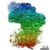
- PDB-3k3q: Crystal Structure of a Llama Antibody complexed with the C. Botul... -
+ Open data

- Basic information
| Entry | Database: PDB / ID: 3k3q | ||||||
|---|---|---|---|---|---|---|---|
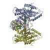
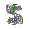
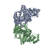
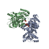
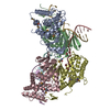
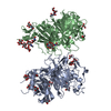
| Title | Crystal Structure of a Llama Antibody complexed with the C. Botulinum Neurotoxin Serotype A Catalytic Domain | ||||||
 Components Components |
| ||||||
 Keywords Keywords | IMMUNE SYSTEM / llama / VHH / antibody / botulinum / neurotoxin / BoNT / Cell junction / Cell membrane / Cytoplasm / Disulfide bond / Hydrolase / Membrane / Metal-binding / Metalloprotease / Pharmaceutical / Protease / Secreted / Synapse / Toxin / Transmembrane / Zinc | ||||||
| Function / homology |  Function and homology information Function and homology informationhost cell junction / bontoxilysin / negative regulation of neurotransmitter secretion / host cell presynaptic membrane / host cell cytoplasmic vesicle / host cell cytosol / transmembrane protein transporter activity / membrane => GO:0016020 / metalloendopeptidase activity / toxin activity ...host cell junction / bontoxilysin / negative regulation of neurotransmitter secretion / host cell presynaptic membrane / host cell cytoplasmic vesicle / host cell cytosol / transmembrane protein transporter activity / membrane => GO:0016020 / metalloendopeptidase activity / toxin activity / proteolysis / extracellular region / zinc ion binding Similarity search - Function | ||||||
| Biological species |   Clostridium botulinum A str. Hall (bacteria) Clostridium botulinum A str. Hall (bacteria) | ||||||
| Method |  X-RAY DIFFRACTION / SYNCHROTRON / MOLECULAR REPLACEMENT / molecular replacement / Resolution: 2.6 Å X-RAY DIFFRACTION / SYNCHROTRON / MOLECULAR REPLACEMENT / molecular replacement / Resolution: 2.6 Å | ||||||
 Authors Authors | Thompson, A.A. / Dong, J. / Marks, J.D. / Stevens, R.C. | ||||||
 Citation Citation | Journal: J.Mol.Biol. / Year: 2010 Title: A Single-Domain Llama Antibody Potently Inhibits the Enzymatic Activity of Botulinum Neurotoxin by Binding to the Non-Catalytic alpha-Exosite Binding Region. Authors: Dong, J. / Thompson, A.A. / Fan, Y. / Lou, J. / Conrad, F. / Ho, M. / Pires-Alves, M. / Wilson, B.A. / Stevens, R.C. / Marks, J.D. | ||||||
| History |
|
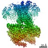
- Structure visualization
| Structure viewer | Molecule: MolmilJmol/JSmol |
|---|
- Downloads & links
-Download
| PDBx/mmCIF format | 3k3q.cif.gz | 123.5 KB | Display | PDBx/mmCIF format |
|---|---|---|---|---|
| PDB format | pdb3k3q.ent.gz | 94 KB | Display | PDB format |
| PDBx/mmJSON format | 3k3q.json.gz | Tree view | PDBx/mmJSON format | |
| Others |  Other downloads Other downloads |
-Validation report
| Arichive directory | https://data.pdbj.org/pub/pdb/validation_reports/k3/3k3qftp://data.pdbj.org/pub/pdb/validation_reports/k3/3k3q | HTTPS FTP |
|---|
-Related structure data
| Similar structure data |
|---|

-Links
 PDBj
PDBj

- Assembly
| Deposited unit | 
| ||||||||
|---|---|---|---|---|---|---|---|---|---|
| 1 | 
| ||||||||
| 2 |
| ||||||||
| Unit cell |
| ||||||||
| Details | Authors state it is possible that a hexamer exists in solution, but this would be in equilibrium with the trimer. |
-Components
| #1: Protein | Mass: 16147.911 Da / Num. of mol.: 1 Source method: isolated from a genetically manipulated source Source: (gene. exp.) |
|---|---|
| #2: Protein | Mass: 28359.986 Da / Num. of mol.: 1 Fragment: N-terminal fragment of BoNT catalytic domain (residues 3-250) Source method: isolated from a genetically manipulated source Source: (gene. exp.) Clostridium botulinum A str. Hall (bacteria)Gene: botA, CBO0806, CLC_0862, neurotoxin catalytic domain / Plasmid: pET15b / Production host: References: UniProt: A5HZZ9, UniProt: P0DPI1*PLUS, bontoxilysin |
| #3: Protein | Mass: 20621.465 Da / Num. of mol.: 1 Fragment: C-terminal fragment of BoNT catalytic domain (residues 251-425) Source method: isolated from a genetically manipulated source Source: (gene. exp.) Clostridium botulinum A str. Hall (bacteria)Gene: botA, CBO0806, CLC_0862, neurotoxin catalytic domain / Plasmid: pET15b / Production host: References: UniProt: A5HZZ9, UniProt: P0DPI1*PLUS, bontoxilysin |
| #4: Chemical | ChemComp-ZN /   Mass: 65.409 Da / Num. of mol.: 1 / Source method: obtained synthetically / Formula: Zn Mass: 65.409 Da / Num. of mol.: 1 / Source method: obtained synthetically / Formula: Zn |
| #5: Water | ChemComp-HOH /  Mass: 18.015 Da / Num. of mol.: 70 / Source method: isolated from a natural source / Formula: H2O Mass: 18.015 Da / Num. of mol.: 70 / Source method: isolated from a natural source / Formula: H2O |
| Has protein modification | Y |
| Sequence details | THERE IS A VAL -> ALA NATURAL VARIANT AT RESIDUE 27. |
-Experimental details
-Experiment
| Experiment | Method: X-RAY DIFFRACTION / Number of used crystals: 1 |
|---|
- Sample preparation
| Crystal | Density Matthews: 2.92 Å3/Da / Density % sol: 57.81 % |
|---|---|
| Crystal grow | Temperature: 293 K / Method: vapor diffusion, sitting drop / pH: 7 Details: 25% ethylene glycol, pH 7.0, VAPOR DIFFUSION, SITTING DROP, temperature 293K |
-Data collection
| Diffraction | Mean temperature: 93 K | |||||||||||||||||||||||||||||||||||||||||||||||||||||||||||||||||||||||||||||||||||||||||||||||||||||||||||||||||||||||||||||||||||||||||||||||||||
|---|---|---|---|---|---|---|---|---|---|---|---|---|---|---|---|---|---|---|---|---|---|---|---|---|---|---|---|---|---|---|---|---|---|---|---|---|---|---|---|---|---|---|---|---|---|---|---|---|---|---|---|---|---|---|---|---|---|---|---|---|---|---|---|---|---|---|---|---|---|---|---|---|---|---|---|---|---|---|---|---|---|---|---|---|---|---|---|---|---|---|---|---|---|---|---|---|---|---|---|---|---|---|---|---|---|---|---|---|---|---|---|---|---|---|---|---|---|---|---|---|---|---|---|---|---|---|---|---|---|---|---|---|---|---|---|---|---|---|---|---|---|---|---|---|---|---|---|---|
| Diffraction source | Source: SYNCHROTRON / Site: APS  / Beamline: 23-ID-B / Wavelength: 1.033 Å / Beamline: 23-ID-B / Wavelength: 1.033 Å | |||||||||||||||||||||||||||||||||||||||||||||||||||||||||||||||||||||||||||||||||||||||||||||||||||||||||||||||||||||||||||||||||||||||||||||||||||
| Detector | Type: MARMOSAIC 300 mm CCD / Detector: CCD / Date: Oct 16, 2008 | |||||||||||||||||||||||||||||||||||||||||||||||||||||||||||||||||||||||||||||||||||||||||||||||||||||||||||||||||||||||||||||||||||||||||||||||||||
| Radiation | Monochromator: Double crystal cryo-cooled Si(111) / Protocol: SINGLE WAVELENGTH / Monochromatic (M) / Laue (L): M / Scattering type: x-ray | |||||||||||||||||||||||||||||||||||||||||||||||||||||||||||||||||||||||||||||||||||||||||||||||||||||||||||||||||||||||||||||||||||||||||||||||||||
| Radiation wavelength | Wavelength: 1.033 Å / Relative weight: 1 | |||||||||||||||||||||||||||||||||||||||||||||||||||||||||||||||||||||||||||||||||||||||||||||||||||||||||||||||||||||||||||||||||||||||||||||||||||
| Reflection | Redundancy: 2.8 % / Av σ(I) over netI: 7.33 / Number: 64075 / Rmerge(I) obs: 0.117 / Χ2: 1 / D res high: 2.6 Å / D res low: 40 Å / Num. obs: 22641 / % possible obs: 96.5 | |||||||||||||||||||||||||||||||||||||||||||||||||||||||||||||||||||||||||||||||||||||||||||||||||||||||||||||||||||||||||||||||||||||||||||||||||||
| Diffraction reflection shell |
| |||||||||||||||||||||||||||||||||||||||||||||||||||||||||||||||||||||||||||||||||||||||||||||||||||||||||||||||||||||||||||||||||||||||||||||||||||
| Reflection | Resolution: 2.6→40 Å / Num. obs: 22641 / % possible obs: 96.5 % / Redundancy: 2.8 % / Biso Wilson estimate: 26.3 Å2 / Rmerge(I) obs: 0.117 / Χ2: 0.995 / Net I/σ(I): 9.5 | |||||||||||||||||||||||||||||||||||||||||||||||||||||||||||||||||||||||||||||||||||||||||||||||||||||||||||||||||||||||||||||||||||||||||||||||||||
| Reflection shell | Resolution: 2.6→2.64 Å / Redundancy: 1.9 % / Rmerge(I) obs: 0.314 / Mean I/σ(I) obs: 2.16 / Num. unique all: 977 / Χ2: 1.003 / % possible all: 83.9 |
-Phasing
| Phasing | Method: molecular replacement |
|---|
- Processing
| Software |
| ||||||||||||||||||||||||
|---|---|---|---|---|---|---|---|---|---|---|---|---|---|---|---|---|---|---|---|---|---|---|---|---|---|
| Refinement | Method to determine structure: MOLECULAR REPLACEMENT / Resolution: 2.6→40 Å / Occupancy max: 1 / Occupancy min: 1 / FOM work R set: 0.823 / Cross valid method: THROUGHOUT / σ(F): 0 / Stereochemistry target values: Engh & Huber
| ||||||||||||||||||||||||
| Solvent computation | Bsol: 20.541 Å2 | ||||||||||||||||||||||||
| Displacement parameters | Biso max: 91.81 Å2 / Biso mean: 41.719 Å2 / Biso min: 3.39 Å2
| ||||||||||||||||||||||||
| Refine analyze |
| ||||||||||||||||||||||||
| Refinement step | Cycle: LAST / Resolution: 2.6→40 Å
| ||||||||||||||||||||||||
| Refine LS restraints |
| ||||||||||||||||||||||||
| Xplor file |
|
