 Movie
Movie Controller

[English] 日本語
 Yorodumi
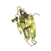
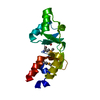
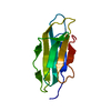
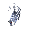
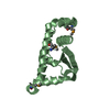
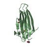
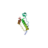
- PDB-3jvc: Crystal Structure of the Lipoprotein_17 domain from Q9PRA0_UREPA ... -
+ Open data

- Basic information
| Entry | Database: PDB / ID: 3jvc | ||||||
|---|---|---|---|---|---|---|---|
| Title | Crystal Structure of the Lipoprotein_17 domain from Q9PRA0_UREPA protein of Ureaplasma parvum. Northeast Structural Genomics Consortium Target UuR17a. | ||||||
 Components Components | Conserved hypothetical membrane lipoprotein | ||||||
 Keywords Keywords | LIPID BINDING PROTEIN / lipoprotein-17 / Q9PRA0 / PF04200 / DUF1976 / UuR17a / NESG. / Structural Genomics / PSI-2 / Protein Structure Initiative / Northeast Structural Genomics Consortium / Lipoprotein | ||||||
| Function / homology |  Function and homology information Function and homology information | ||||||
| Biological species |  Ureaplasma parvum serovar 3 str. ATCC 700970 (bacteria) Ureaplasma parvum serovar 3 str. ATCC 700970 (bacteria) | ||||||
| Method |  X-RAY DIFFRACTION / SYNCHROTRON / SAD / Resolution: 2.69 Å X-RAY DIFFRACTION / SYNCHROTRON / SAD / Resolution: 2.69 Å | ||||||
 Authors Authors | Vorobiev, S. / Neely, H. / Lee, D. / Ciccosanti, C. / Mao, L. / Xiao, R. / Acton, T.B. / Montelione, G.T. / Tong, L. / Hunt, J.F. / Northeast Structural Genomics Consortium (NESG) | ||||||
 Citation Citation | Journal: To be Published Title: Crystal Structure of the Lipoprotein_17 domain from Q9PRA0_UREPA protein of Ureaplasma parvum. Authors: Vorobiev, S. / Neely, H. / Lee, D. / Ciccosanti, C. / Mao, L. / Xiao, R. / Acton, T.B. / Montelione, G.T. / Tong, L. / Hunt, J.F. | ||||||
| History |
|
- Structure visualization
| Structure viewer | Molecule: MolmilJmol/JSmol |
|---|
- Downloads & links
-Download
| PDBx/mmCIF format | 3jvc.cif.gz | 77.9 KB | Display | PDBx/mmCIF format |
|---|---|---|---|---|
| PDB format | pdb3jvc.ent.gz | 60.1 KB | Display | PDB format |
| PDBx/mmJSON format | 3jvc.json.gz | Tree view | PDBx/mmJSON format | |
| Others |  Other downloads Other downloads |
-Validation report
| Arichive directory | https://data.pdbj.org/pub/pdb/validation_reports/jv/3jvcftp://data.pdbj.org/pub/pdb/validation_reports/jv/3jvc | HTTPS FTP |
|---|
-Related structure data
| Similar structure data | |
|---|---|
| Other databases |


-Links
 PDBj
PDBj


- Assembly
| Deposited unit | 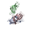
| ||||||||
|---|---|---|---|---|---|---|---|---|---|
| 1 | 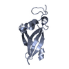
| ||||||||
| 2 | 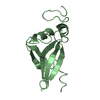
| ||||||||
| 3 | 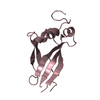
| ||||||||
| Unit cell |
| ||||||||
| Details | monomer according to gel-filtration |
-Components
| #1: Protein | Mass: 14782.609 Da / Num. of mol.: 3 Source method: isolated from a genetically manipulated source Source: (gene. exp.) Ureaplasma parvum serovar 3 str. ATCC 700970 (bacteria)Strain: ATCC 700970 / Serovar 3 / Gene: UU045 / Plasmid: pET 21-23C / Production host: #2: Water | ChemComp-HOH / |  Mass: 18.015 Da / Num. of mol.: 40 / Source method: isolated from a natural source / Formula: H2O Mass: 18.015 Da / Num. of mol.: 40 / Source method: isolated from a natural source / Formula: H2O |
|---|
-Experimental details
-Experiment
| Experiment | Method: X-RAY DIFFRACTION / Number of used crystals: 1 |
|---|
- Sample preparation
| Crystal | Density Matthews: 2.35 Å3/Da / Density % sol: 47.65 % |
|---|---|
| Crystal grow | Temperature: 291 K / Method: microbatch under paraffin oil / pH: 7.5 Details: 24% PEG 20000, 0.1M RbCl, 0.1M HEPES, pH 7.5, microbatch under paraffin oil, temperature 291K |
-Data collection
| Diffraction | Mean temperature: 100 K |
|---|---|
| Diffraction source | Source: SYNCHROTRON / Site: NSLS  / Beamline: X4A / Wavelength: 0.97914 Å / Beamline: X4A / Wavelength: 0.97914 Å |
| Detector | Type: ADSC QUANTUM 4 / Detector: CCD / Date: Aug 19, 2009 |
| Radiation | Protocol: SINGLE WAVELENGTH / Monochromatic (M) / Laue (L): M / Scattering type: x-ray |
| Radiation wavelength | Wavelength: 0.97914 Å / Relative weight: 1 |
| Reflection | Resolution: 2.69→50 Å / Num. all: 22432 / Num. obs: 22095 / % possible obs: 98.5 % / Observed criterion σ(F): 0 / Observed criterion σ(I): 0 / Redundancy: 11.9 % / Biso Wilson estimate: 48.5 Å2 / Rmerge(I) obs: 0.061 / Net I/σ(I): 52.2 |
| Reflection shell | Resolution: 2.69→2.79 Å / Redundancy: 9.8 % / Rmerge(I) obs: 0.212 / Mean I/σ(I) obs: 14.2 / Num. unique all: 2184 / % possible all: 94 |
- Processing
| Software |
| ||||||||||||||||||||||||
|---|---|---|---|---|---|---|---|---|---|---|---|---|---|---|---|---|---|---|---|---|---|---|---|---|---|
| Refinement | Method to determine structure: SAD / Resolution: 2.69→38.42 Å / Rfactor Rfree error: 0.008 / Data cutoff high absF: 161344.13 / Data cutoff low absF: 0 / Isotropic thermal model: RESTRAINED / Cross valid method: THROUGHOUT / σ(F): 0 / Stereochemistry target values: Engh & Huber / Details: BULK SOLVENT MODEL USED
| ||||||||||||||||||||||||
| Solvent computation | Solvent model: FLAT MODEL / Bsol: 37.4848 Å2 / ksol: 0.3 e/Å3 | ||||||||||||||||||||||||
| Displacement parameters | Biso mean: 56.5 Å2
| ||||||||||||||||||||||||
| Refine analyze |
| ||||||||||||||||||||||||
| Refinement step | Cycle: LAST / Resolution: 2.69→38.42 Å
| ||||||||||||||||||||||||
| Refine LS restraints |
| ||||||||||||||||||||||||
| Refine LS restraints NCS | NCS model details: NONE | ||||||||||||||||||||||||
| LS refinement shell | Resolution: 2.69→2.86 Å / Rfactor Rfree error: 0.028 / Total num. of bins used: 6
| ||||||||||||||||||||||||
| Xplor file |
|
