 Movie
Movie Controller

[English] 日本語
 Yorodumi
- PDB-3jug: Crystal structure of endo-beta-1,4-mannanase from the alkaliphili... -
+ Open data

- Basic information
| Entry | Database: PDB / ID: 3jug | ||||||
|---|---|---|---|---|---|---|---|
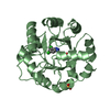
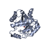
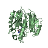
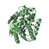
| Title | Crystal structure of endo-beta-1,4-mannanase from the alkaliphilic Bacillus sp. N16-5 | ||||||
 Components Components | Beta-mannanase | ||||||
 Keywords Keywords | HYDROLASE / TIM-BARREL / Glycosidase | ||||||
| Function / homology |  Function and homology information Function and homology informationbeta-mannosidase activity / mannan endo-1,4-beta-mannosidase / mannan endo-1,4-beta-mannosidase activity / glucan catabolic process / membrane Similarity search - Function | ||||||
| Biological species |  | ||||||
| Method |  X-RAY DIFFRACTION / MOLECULAR REPLACEMENT / Resolution: 1.6 Å X-RAY DIFFRACTION / MOLECULAR REPLACEMENT / Resolution: 1.6 Å | ||||||
 Authors Authors | Zhao, Y. / Zhang, Y. / Xue, Y. | ||||||
 Citation Citation | Journal: To be Published Title: Structure Analysis of Alkaline beta-mannanase from Alkaliphilic Bacillus sp. N16-5 Implications for Adaptation to Alkaline Conditions Authors: Zhao, Y. / Zhang, Y. / Cao, Y. / Qi, J. / Xue, Y. / Gao, F. / Peng, H. / Gao, G.F. / Ma, Y. | ||||||
| History |
|
- Structure visualization
| Structure viewer | Molecule: MolmilJmol/JSmol |
|---|
- Downloads & links
-Download
| PDBx/mmCIF format | 3jug.cif.gz | 150.7 KB | Display | PDBx/mmCIF format |
|---|---|---|---|---|
| PDB format | pdb3jug.ent.gz | 115.6 KB | Display | PDB format |
| PDBx/mmJSON format | 3jug.json.gz | Tree view | PDBx/mmJSON format | |
| Others |  Other downloads Other downloads |
-Validation report
| Summary document | 3jug_validation.pdf.gz | 442.1 KB | Display | wwPDB validaton report |
|---|---|---|---|---|
| Full document | 3jug_full_validation.pdf.gz | 444.5 KB | Display | |
| Data in XML | 3jug_validation.xml.gz | 19.3 KB | Display | |
| Data in CIF | 3jug_validation.cif.gz | 30.9 KB | Display | |
| Arichive directory | https://data.pdbj.org/pub/pdb/validation_reports/ju/3jugftp://data.pdbj.org/pub/pdb/validation_reports/ju/3jug | HTTPS FTP |
-Related structure data
| Related structure data |  1wkyS S: Starting model for refinement |
|---|---|
| Similar structure data |


-Links
 PDBj
PDBj- Assembly
| Deposited unit | 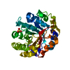
| ||||||||
|---|---|---|---|---|---|---|---|---|---|
| 1 |
| ||||||||
| Unit cell |
|
-Components
| #1: Protein | Mass: 38168.770 Da / Num. of mol.: 1 / Fragment: endo-beta-mannanase, UNP residues 32-330 Source method: isolated from a genetically manipulated source Source: (gene. exp.) References: UniProt: Q5YEX6, mannan endo-1,4-beta-mannosidase |
|---|---|
| #2: Chemical | ChemComp-SO4 /   Mass: 96.063 Da / Num. of mol.: 1 / Source method: obtained synthetically / Formula: SO4 Mass: 96.063 Da / Num. of mol.: 1 / Source method: obtained synthetically / Formula: SO4 |
| #3: Water | ChemComp-HOH /  Mass: 18.015 Da / Num. of mol.: 589 / Source method: isolated from a natural source / Formula: H2O Mass: 18.015 Da / Num. of mol.: 589 / Source method: isolated from a natural source / Formula: H2O |
-Experimental details
-Experiment
| Experiment | Method: X-RAY DIFFRACTION / Number of used crystals: 1 |
|---|
- Sample preparation
| Crystal | Density Matthews: 2.04 Å3/Da / Density % sol: 39.71 % |
|---|---|
| Crystal grow | Temperature: 298 K / Method: vapor diffusion, hanging drop / pH: 5.6 Details: 0.5M Ammonium Sulphate, 0.1M sodium citrate, 1.2M Lithium Sulfate, pH 5.6, VAPOR DIFFUSION, HANGING DROP, temperature 298K |
-Data collection
| Diffraction | Mean temperature: 100 K |
|---|---|
| Diffraction source | Source: ROTATING ANODE / Type: RIGAKU MICROMAX-007 / Wavelength: 1.5418 Å |
| Detector | Type: RIGAKU RAXIS IV / Detector: IMAGE PLATE / Date: Jul 20, 2005 / Details: mirrors |
| Radiation | Monochromator: GRAPHITE / Protocol: SINGLE WAVELENGTH / Monochromatic (M) / Laue (L): M / Scattering type: x-ray |
| Radiation wavelength | Wavelength: 1.5418 Å / Relative weight: 1 |
| Reflection | Resolution: 1.6→63.29 Å / Num. obs: 38945 / % possible obs: 92.74 % / Observed criterion σ(F): 2 / Observed criterion σ(I): 2 / Redundancy: 4.68 % / Biso Wilson estimate: 6.94 Å2 / Rsym value: 0.0764 |
| Reflection shell | Resolution: 1.6→1.71 Å / Redundancy: 1.54 % / Num. unique all: 4841 / % possible all: 65.44 |
- Processing
| Software |
| ||||||||||||||||||||||||||||||||||||||||||||||||||||||||||||||||||||||||||||||||||||||||||||||||||
|---|---|---|---|---|---|---|---|---|---|---|---|---|---|---|---|---|---|---|---|---|---|---|---|---|---|---|---|---|---|---|---|---|---|---|---|---|---|---|---|---|---|---|---|---|---|---|---|---|---|---|---|---|---|---|---|---|---|---|---|---|---|---|---|---|---|---|---|---|---|---|---|---|---|---|---|---|---|---|---|---|---|---|---|---|---|---|---|---|---|---|---|---|---|---|---|---|---|---|---|
| Refinement | Method to determine structure: MOLECULAR REPLACEMENT Starting model: PDB ENTRY 1WKY Resolution: 1.6→43.176 Å / FOM work R set: 0.86 / SU ML: 0.19 / σ(F): 0.13 / Phase error: 20.77 / Stereochemistry target values: ML
| ||||||||||||||||||||||||||||||||||||||||||||||||||||||||||||||||||||||||||||||||||||||||||||||||||
| Solvent computation | Shrinkage radii: 0.9 Å / VDW probe radii: 1.11 Å / Solvent model: FLAT BULK SOLVENT MODEL / Bsol: 51.519 Å2 / ksol: 0.352 e/Å3 | ||||||||||||||||||||||||||||||||||||||||||||||||||||||||||||||||||||||||||||||||||||||||||||||||||
| Displacement parameters | Biso mean: 12.388 Å2
| ||||||||||||||||||||||||||||||||||||||||||||||||||||||||||||||||||||||||||||||||||||||||||||||||||
| Refinement step | Cycle: LAST / Resolution: 1.6→43.176 Å
| ||||||||||||||||||||||||||||||||||||||||||||||||||||||||||||||||||||||||||||||||||||||||||||||||||
| Refine LS restraints |
| ||||||||||||||||||||||||||||||||||||||||||||||||||||||||||||||||||||||||||||||||||||||||||||||||||
| LS refinement shell |
| ||||||||||||||||||||||||||||||||||||||||||||||||||||||||||||||||||||||||||||||||||||||||||||||||||
| Refinement TLS params. | Method: refined / Origin x: 17.1848 Å / Origin y: -1.5875 Å / Origin z: -11.9158 Å
| ||||||||||||||||||||||||||||||||||||||||||||||||||||||||||||||||||||||||||||||||||||||||||||||||||
| Refinement TLS group |
|
