 Movie
Movie Controller

[English] 日本語
 Yorodumi
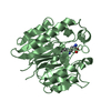
- PDB-3i4a: Crystal structure of dimethylarginine dimethylaminohydrolase-1 (D... -
+ Open data

- Basic information
| Entry | Database: PDB / ID: 3i4a | ||||||
|---|---|---|---|---|---|---|---|
| Title | Crystal structure of dimethylarginine dimethylaminohydrolase-1 (DDAH-1) in complex with N5-(1-iminopropyl)-L-ornithine | ||||||
 Components Components | N(G),N(G)-dimethylarginine dimethylaminohydrolase 1 | ||||||
 Keywords Keywords | HYDROLASE / DDAH / NITRIC OXIDE SYNTHASE REGULATION / Metal-binding | ||||||
| Function / homology |  Function and homology information Function and homology informationdimethylargininase / dimethylargininase activity / : / negative regulation of cellular response to hypoxia / arginine metabolic process / regulation of systemic arterial blood pressure / negative regulation of vascular permeability / amino acid binding / catalytic activity / : ...dimethylargininase / dimethylargininase activity / : / negative regulation of cellular response to hypoxia / arginine metabolic process / regulation of systemic arterial blood pressure / negative regulation of vascular permeability / amino acid binding / catalytic activity / : / L-arginine catabolic process / nitric oxide metabolic process / eNOS activation / positive regulation of angiogenesis / positive regulation of nitric oxide biosynthetic process / negative regulation of cell population proliferation / extracellular exosome / metal ion binding / cytosol Similarity search - Function | ||||||
| Biological species |  Homo sapiens (human) Homo sapiens (human) | ||||||
| Method |  X-RAY DIFFRACTION / MOLECULAR REPLACEMENT / Resolution: 1.898 Å X-RAY DIFFRACTION / MOLECULAR REPLACEMENT / Resolution: 1.898 Å | ||||||
 Authors Authors | Monzingo, A.F. / Wang, Y. / Hu, S. / Schaller, T.H. / Fast, W. / Robertus, J.D. | ||||||
 Citation Citation | Journal: Biochemistry / Year: 2009 Title: Developing dual and specific inhibitors of dimethylarginine dimethylaminohydrolase-1 and nitric oxide synthase: toward a targeted polypharmacology to control nitric oxide. Authors: Wang, Y. / Monzingo, A.F. / Hu, S. / Schaller, T.H. / Robertus, J.D. / Fast, W. | ||||||
| History |
|
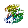
- Structure visualization
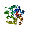
| Structure viewer | Molecule: MolmilJmol/JSmol |
|---|
- Downloads & links
-Download
| PDBx/mmCIF format | 3i4a.cif.gz | 119 KB | Display | PDBx/mmCIF format |
|---|---|---|---|---|
| PDB format | pdb3i4a.ent.gz | 90.6 KB | Display | PDB format |
| PDBx/mmJSON format | 3i4a.json.gz | Tree view | PDBx/mmJSON format | |
| Others |  Other downloads Other downloads |
-Validation report
| Arichive directory | https://data.pdbj.org/pub/pdb/validation_reports/i4/3i4aftp://data.pdbj.org/pub/pdb/validation_reports/i4/3i4a | HTTPS FTP |
|---|
-Related structure data
| Related structure data |  3i2eSC S: Starting model for refinement C: citing same article ( |
|---|---|
| Similar structure data |


-Links
 PDBj
PDBj


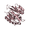
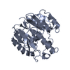
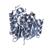
- Assembly
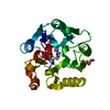
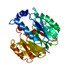
| Deposited unit | 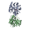
| |||||||||||||||||||||||||||
|---|---|---|---|---|---|---|---|---|---|---|---|---|---|---|---|---|---|---|---|---|---|---|---|---|---|---|---|---|
| 1 | 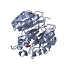
| |||||||||||||||||||||||||||
| 2 | 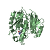
| |||||||||||||||||||||||||||
| Unit cell |
| |||||||||||||||||||||||||||
| Noncrystallographic symmetry (NCS) | NCS domain:
NCS domain segments:
NCS oper: (Code: given Matrix: (0.999324, -0.036751, 0.0006), Vector: |
-Components
| #1: Protein | Mass: 33623.492 Da / Num. of mol.: 2 Source method: isolated from a genetically manipulated source Source: (gene. exp.) Homo sapiens (human) / Gene: DDAH, DDAH1 / Plasmid: PET28A-HDDAH-1 / Production host:  #2: Chemical | 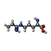  Mass: 187.239 Da / Num. of mol.: 2 / Source method: obtained synthetically / Formula: C8H17N3O2 Mass: 187.239 Da / Num. of mol.: 2 / Source method: obtained synthetically / Formula: C8H17N3O2#3: Water | ChemComp-HOH / |  Mass: 18.015 Da / Num. of mol.: 99 / Source method: isolated from a natural source / Formula: H2O Mass: 18.015 Da / Num. of mol.: 99 / Source method: isolated from a natural source / Formula: H2OHas protein modification | Y | |
|---|
-Experimental details
-Experiment
| Experiment | Method: X-RAY DIFFRACTION / Number of used crystals: 1 |
|---|
- Sample preparation
| Crystal | Density Matthews: 2.1 Å3/Da / Density % sol: 41.31 % |
|---|---|
| Crystal grow | Temperature: 277 K / Method: vapor diffusion, hanging drop / pH: 8 Details: 25% (w/v) PEG 6000, 0.1 M Tris-HCl, pH 8.0, VAPOR DIFFUSION, HANGING DROP, temperature 277K |
-Data collection
| Diffraction | Mean temperature: 100 K |
|---|---|
| Diffraction source | Source: ROTATING ANODE / Type: RIGAKU MICROMAX-007 HF / Wavelength: 1.5418 Å |
| Detector | Type: RIGAKU RAXIS HTC / Detector: IMAGE PLATE / Date: May 21, 2009 / Details: VariMax High Flux Optics |
| Radiation | Monochromator: VariMax High Flux Optics / Protocol: SINGLE WAVELENGTH / Monochromatic (M) / Laue (L): M / Scattering type: x-ray |
| Radiation wavelength | Wavelength: 1.5418 Å / Relative weight: 1 |
| Reflection twin | Operator: h,-k,-l / Fraction: 0.486 |
| Reflection | Resolution: 1.898→25.12 Å / Num. obs: 42222 / % possible obs: 96.1 % / Observed criterion σ(I): 0 / Redundancy: 3.7 % / Rmerge(I) obs: 0.065 / Χ2: 1.078 / Net I/σ(I): 10.5 |
| Reflection shell | Resolution: 1.898→1.93 Å / Redundancy: 3.6 % / Rmerge(I) obs: 0.391 / Num. unique all: 2048 / Χ2: 0.6 / % possible all: 94.7 |
- Processing
| Software |
| ||||||||||||||||||||||||||||||||||||||||||||||||||||||||||||||||||||||||||||||||||||||||||||||||||||||||||||||||||||||||||||||
|---|---|---|---|---|---|---|---|---|---|---|---|---|---|---|---|---|---|---|---|---|---|---|---|---|---|---|---|---|---|---|---|---|---|---|---|---|---|---|---|---|---|---|---|---|---|---|---|---|---|---|---|---|---|---|---|---|---|---|---|---|---|---|---|---|---|---|---|---|---|---|---|---|---|---|---|---|---|---|---|---|---|---|---|---|---|---|---|---|---|---|---|---|---|---|---|---|---|---|---|---|---|---|---|---|---|---|---|---|---|---|---|---|---|---|---|---|---|---|---|---|---|---|---|---|---|---|---|
| Refinement | Method to determine structure: MOLECULAR REPLACEMENT Starting model: PDB entry 3I2E Resolution: 1.898→25.12 Å / Occupancy max: 1 / Occupancy min: 1 / Isotropic thermal model: isotropic / Cross valid method: THROUGHOUT / σ(F): 0 / Stereochemistry target values: Engh & Huber / Details: detwinning algorithm used during refinement
| ||||||||||||||||||||||||||||||||||||||||||||||||||||||||||||||||||||||||||||||||||||||||||||||||||||||||||||||||||||||||||||||
| Solvent computation | Shrinkage radii: 0.9 Å / VDW probe radii: 1.11 Å / Solvent model: FLAT BULK SOLVENT MODEL / Bsol: 29.55 Å2 / ksol: 0.302 e/Å3 | ||||||||||||||||||||||||||||||||||||||||||||||||||||||||||||||||||||||||||||||||||||||||||||||||||||||||||||||||||||||||||||||
| Displacement parameters | Biso max: 60.25 Å2 / Biso mean: 30.402 Å2 / Biso min: 8.34 Å2
| ||||||||||||||||||||||||||||||||||||||||||||||||||||||||||||||||||||||||||||||||||||||||||||||||||||||||||||||||||||||||||||||
| Refinement step | Cycle: LAST / Resolution: 1.898→25.12 Å
| ||||||||||||||||||||||||||||||||||||||||||||||||||||||||||||||||||||||||||||||||||||||||||||||||||||||||||||||||||||||||||||||
| Refine LS restraints |
| ||||||||||||||||||||||||||||||||||||||||||||||||||||||||||||||||||||||||||||||||||||||||||||||||||||||||||||||||||||||||||||||
| Refine LS restraints NCS |
| ||||||||||||||||||||||||||||||||||||||||||||||||||||||||||||||||||||||||||||||||||||||||||||||||||||||||||||||||||||||||||||||
| LS refinement shell | Refine-ID: X-RAY DIFFRACTION / Total num. of bins used: 20 / % reflection obs: 98 %
|
