 Movie
Movie Controller

[English] 日本語
 Yorodumi
- PDB-3jqj: Crystal structure of the molybdenum cofactor biosynthesis protein... -
+ Open data

- Basic information
| Entry | Database: PDB / ID: 3jqj | ||||||
|---|---|---|---|---|---|---|---|
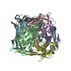
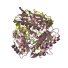
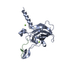
| Title | Crystal structure of the molybdenum cofactor biosynthesis protein C (TTHA1789) from Thermus Theromophilus HB8 | ||||||
 Components Components | Molybdenum cofactor biosynthesis protein C | ||||||
 Keywords Keywords | BIOSYNTHETIC PROTEIN / MOAC / MOLYBDENUM COFACTOR (MOCO) / MOCO BIOSYNTHESIS / STRUCTURAL GENOMICS / NPPSFA / NATIONAL PROJECT ON PROTEIN STRUCTURAL AND FUNCTIONAL ANALYSES / RIKEN STRUCTURAL GENOMICS/PROTEOMICS INITIATIVE / RSGI / Molybdenum cofactor biosynthesis | ||||||
| Function / homology |  Function and homology information Function and homology informationcyclic pyranopterin monophosphate synthase / cyclic pyranopterin monophosphate synthase activity / Mo-molybdopterin cofactor biosynthetic process / GTP binding Similarity search - Function | ||||||
| Biological species |   Thermus thermophilus (bacteria) Thermus thermophilus (bacteria) | ||||||
| Method |  X-RAY DIFFRACTION / SYNCHROTRON / MOLECULAR REPLACEMENT / Resolution: 1.9 Å X-RAY DIFFRACTION / SYNCHROTRON / MOLECULAR REPLACEMENT / Resolution: 1.9 Å | ||||||
 Authors Authors | Kanaujia, S.P. / Jeyakanthan, J. / Nakagawa, N. / Sekar, K. / Baba, S. / Ebihara, A. / Kuramitsu, S. / Shinkai, A. / Shiro, Y. / Yokoyama, S. / RIKEN Structural Genomics/Proteomics Initiative (RSGI) | ||||||
 Citation Citation | Journal: Acta Crystallogr.,Sect.D / Year: 2010 Title: Structures of apo and GTP-bound molybdenum cofactor biosynthesis protein MoaC from Thermus thermophilus HB8 Authors: Kanaujia, S.P. / Jeyakanthan, J. / Nakagawa, N. / Balasubramaniam, S. / Shinkai, A. / Kuramitsu, S. / Yokoyama, S. / Sekar, K. | ||||||
| History |
|
- Structure visualization
| Structure viewer | Molecule: MolmilJmol/JSmol |
|---|
- Downloads & links
-Download
| PDBx/mmCIF format | 3jqj.cif.gz | 361.1 KB | Display | PDBx/mmCIF format |
|---|---|---|---|---|
| PDB format | pdb3jqj.ent.gz | 294.6 KB | Display | PDB format |
| PDBx/mmJSON format | 3jqj.json.gz | Tree view | PDBx/mmJSON format | |
| Others |  Other downloads Other downloads |
-Validation report
| Arichive directory | https://data.pdbj.org/pub/pdb/validation_reports/jq/3jqjftp://data.pdbj.org/pub/pdb/validation_reports/jq/3jqj | HTTPS FTP |
|---|
-Related structure data
| Related structure data |  3jqkC  3jqmC 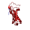 1ekrS C: citing same article ( S: Starting model for refinement |
|---|---|
| Similar structure data | |
| Other databases |


-Links
 PDBj
PDBj- Assembly
| Deposited unit | 
| ||||||||
|---|---|---|---|---|---|---|---|---|---|
| 1 | 
| ||||||||
| 2 | 
| ||||||||
| Unit cell |
|
-Components
| #1: Protein | Mass: 16946.742 Da / Num. of mol.: 12 Source method: isolated from a genetically manipulated source Source: (gene. exp.) Thermus thermophilus (bacteria) / Strain: HB8 / Gene: TTHA1789 / Plasmid: PET11A / Production host: #2: Chemical | ChemComp-PO4 /   Mass: 94.971 Da / Num. of mol.: 12 / Source method: obtained synthetically / Formula: PO4 Mass: 94.971 Da / Num. of mol.: 12 / Source method: obtained synthetically / Formula: PO4#3: Chemical | ChemComp-PGR /   Mass: 76.094 Da / Num. of mol.: 10 / Source method: obtained synthetically / Formula: C3H8O2 Mass: 76.094 Da / Num. of mol.: 10 / Source method: obtained synthetically / Formula: C3H8O2#4: Chemical | ChemComp-GOL /   Mass: 92.094 Da / Num. of mol.: 12 / Source method: obtained synthetically / Formula: C3H8O3 Mass: 92.094 Da / Num. of mol.: 12 / Source method: obtained synthetically / Formula: C3H8O3#5: Water | ChemComp-HOH / |  Mass: 18.015 Da / Num. of mol.: 1181 / Source method: isolated from a natural source / Formula: H2O Mass: 18.015 Da / Num. of mol.: 1181 / Source method: isolated from a natural source / Formula: H2O |
|---|
-Experimental details
-Experiment
| Experiment | Method: X-RAY DIFFRACTION / Number of used crystals: 1 |
|---|
- Sample preparation
| Crystal | Density Matthews: 1.95 Å3/Da / Density % sol: 36.88 % |
|---|---|
| Crystal grow | Temperature: 293 K / Method: vapor diffusion, sitting drop / pH: 4.2 Details: 25%, 1,2-PROPANEDIOL, 5% PEG 3000, 0.1M PHOSPHATE-CITRATE PH 4.2, 10% GLYCEROL,, VAPOR DIFFUSION, SITTING DROP, temperature 293K |
-Data collection
| Diffraction source | Source: SYNCHROTRON / Site: SPring-8  / Beamline: BL26B2 / Wavelength: 1 Å / Beamline: BL26B2 / Wavelength: 1 Å |
|---|---|
| Detector | Type: RIGAKU / Detector: IMAGE PLATE / Date: Jul 7, 2006 / Details: RH COATED BENT-CYLINDRICAL MIRROR |
| Radiation | Monochromator: SI-111 DOUBLE CRYSTAL / Protocol: SINGLE WAVELENGTH / Monochromatic (M) / Laue (L): M / Scattering type: x-ray |
| Radiation wavelength | Wavelength: 1 Å / Relative weight: 1 |
| Reflection | Resolution: 1.9→50 Å / Num. obs: 121501 / % possible obs: 99.9 % / Biso Wilson estimate: 12.9 Å2 / Rmerge(I) obs: 0.045 / Rsym value: 0.042 |
| Reflection shell | Resolution: 1.9→1.97 Å / Rmerge(I) obs: 0.172 / Num. unique all: 12072 / Rsym value: 0.174 / % possible all: 99.7 |
- Processing
| Software |
| ||||||||||||||||||||
|---|---|---|---|---|---|---|---|---|---|---|---|---|---|---|---|---|---|---|---|---|---|
| Refinement | Method to determine structure: MOLECULAR REPLACEMENT Starting model: PDB ENTRY 1EKR Resolution: 1.9→49.66 Å / Rfactor Rfree error: 0.003 / Data cutoff high absF: 604951.65 / Data cutoff low absF: 0 / Isotropic thermal model: RESTRAINED / Cross valid method: THROUGHOUT / σ(F): 0 / Stereochemistry target values: Engh & Huber / Details: BULK SOLVENT MODEL USED
| ||||||||||||||||||||
| Solvent computation | Solvent model: FLAT MODEL / Bsol: 68.0318 Å2 / ksol: 0.4 e/Å3 | ||||||||||||||||||||
| Displacement parameters | Biso mean: 24.5 Å2
| ||||||||||||||||||||
| Refine analyze |
| ||||||||||||||||||||
| Refinement step | Cycle: LAST / Resolution: 1.9→49.66 Å
| ||||||||||||||||||||
| Refine LS restraints |
| ||||||||||||||||||||
| LS refinement shell | Resolution: 1.9→2.02 Å / Rfactor Rfree error: 0.008 / Total num. of bins used: 6
| ||||||||||||||||||||
| Xplor file |
|
