 Movie
Movie Controller

[English] 日本語
 Yorodumi
- PDB-2ohd: Crystal structure of hypothetical molybdenum cofactor biosynthesi... -
+ Open data

- Basic information
| Entry | Database: PDB / ID: 2ohd | ||||||
|---|---|---|---|---|---|---|---|
| Title | Crystal structure of hypothetical molybdenum cofactor biosynthesis protein C from Sulfolobus tokodaii | ||||||
 Components Components | Probable molybdenum cofactor biosynthesis protein C | ||||||
 Keywords Keywords | BIOSYNTHETIC PROTEIN / alpha + beta | ||||||
| Function / homology |  Function and homology information Function and homology informationGTP 3',8'-cyclase activity / cyclic pyranopterin monophosphate synthase / cyclic pyranopterin monophosphate synthase activity / Mo-molybdopterin cofactor biosynthetic process Similarity search - Function | ||||||
| Biological species |   Sulfolobus tokodaii str. 7 (archaea) Sulfolobus tokodaii str. 7 (archaea) | ||||||
| Method |  X-RAY DIFFRACTION / SYNCHROTRON / MOLECULAR REPLACEMENT / Resolution: 2.2 Å X-RAY DIFFRACTION / SYNCHROTRON / MOLECULAR REPLACEMENT / Resolution: 2.2 Å | ||||||
 Authors Authors | Yoshida, H. / Yamada, M. / Kuramitsu, S. / Kamitori, S. | ||||||
 Citation Citation | Journal: Acta Crystallogr.,Sect.F / Year: 2008 Title: Structure of a putative molybdenum-cofactor biosynthesis protein C (MoaC) from Sulfolobus tokodaii (ST0472) Authors: Yoshida, H. / Yamada, M. / Kuramitsu, S. / Kamitori, S. | ||||||
| History |
|
- Structure visualization
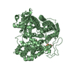
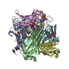
| Structure viewer | Molecule: MolmilJmol/JSmol |
|---|
- Downloads & links
-Download
| PDBx/mmCIF format | 2ohd.cif.gz | 179.5 KB | Display | PDBx/mmCIF format |
|---|---|---|---|---|
| PDB format | pdb2ohd.ent.gz | 144.9 KB | Display | PDB format |
| PDBx/mmJSON format | 2ohd.json.gz | Tree view | PDBx/mmJSON format | |
| Others |  Other downloads Other downloads |
-Validation report
| Arichive directory | https://data.pdbj.org/pub/pdb/validation_reports/oh/2ohdftp://data.pdbj.org/pub/pdb/validation_reports/oh/2ohd | HTTPS FTP |
|---|
-Related structure data
| Related structure data | 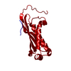 1ekrS S: Starting model for refinement |
|---|---|
| Similar structure data |


-Links
 PDBj
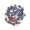
PDBj- Assembly
| Deposited unit | 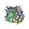
| ||||||||
|---|---|---|---|---|---|---|---|---|---|
| 1 |
| ||||||||
| Unit cell |
|
-Components
| #1: Protein | Mass: 17152.123 Da / Num. of mol.: 6 Source method: isolated from a genetically manipulated source Source: (gene. exp.) Sulfolobus tokodaii str. 7 (archaea) / Species: Sulfolobus tokodaii / Strain: strain 7 / Gene: ST0472 / Production host:  #2: Water | ChemComp-HOH / |  Mass: 18.015 Da / Num. of mol.: 443 / Source method: isolated from a natural source / Formula: H2O Mass: 18.015 Da / Num. of mol.: 443 / Source method: isolated from a natural source / Formula: H2O |
|---|
-Experimental details
-Experiment
| Experiment | Method: X-RAY DIFFRACTION / Number of used crystals: 1 |
|---|
- Sample preparation
| Crystal | Density Matthews: 2.34 Å3/Da / Density % sol: 47.43 % |
|---|---|
| Crystal grow | Temperature: 298 K / Method: vapor diffusion, hanging drop / pH: 7.5 Details: 18% PEG 3000, 0.1M HEPES, 0.2M magnesium chloride , pH 7.5, VAPOR DIFFUSION, HANGING DROP, temperature 298K |
-Data collection
| Diffraction | Mean temperature: 100 K |
|---|---|
| Diffraction source | Source: SYNCHROTRON / Site: Photon Factory  / Beamline: BL-6A / Wavelength: 0.978 Å / Beamline: BL-6A / Wavelength: 0.978 Å |
| Detector | Type: ADSC QUANTUM 4 / Detector: CCD / Date: Dec 1, 2006 |
| Radiation | Monochromator: GRAPHITE / Protocol: SINGLE WAVELENGTH / Monochromatic (M) / Laue (L): M / Scattering type: x-ray |
| Radiation wavelength | Wavelength: 0.978 Å / Relative weight: 1 |
| Reflection | Resolution: 2.2→50 Å / Num. all: 48362 / Num. obs: 48362 / % possible obs: 99.9 % / Observed criterion σ(F): 0 / Observed criterion σ(I): 0 / Redundancy: 3.4 % / Biso Wilson estimate: 15.2 Å2 / Rmerge(I) obs: 0.055 / Net I/σ(I): 11.3 |
| Reflection shell | Resolution: 2.2→2.28 Å / Rmerge(I) obs: 0.268 / % possible all: 99.6 |
- Processing
| Software |
| |||||||||||||||||||||||||
|---|---|---|---|---|---|---|---|---|---|---|---|---|---|---|---|---|---|---|---|---|---|---|---|---|---|---|
| Refinement | Method to determine structure: MOLECULAR REPLACEMENT Starting model: PDB ENTRY 1EKR Resolution: 2.2→45.78 Å / Rfactor Rfree error: 0.004 / Data cutoff high absF: 52270.57 / Data cutoff low absF: 0 / Isotropic thermal model: RESTRAINED / Cross valid method: THROUGHOUT / σ(F): 0 / Stereochemistry target values: Engh & Huber
| |||||||||||||||||||||||||
| Solvent computation | Solvent model: FLAT MODEL / Bsol: 49.5666 Å2 / ksol: 0.373335 e/Å3 | |||||||||||||||||||||||||
| Displacement parameters | Biso mean: 29.4 Å2
| |||||||||||||||||||||||||
| Refine analyze |
| |||||||||||||||||||||||||
| Refinement step | Cycle: LAST / Resolution: 2.2→45.78 Å
| |||||||||||||||||||||||||
| Refine LS restraints |
| |||||||||||||||||||||||||
| LS refinement shell | Resolution: 2.2→2.34 Å / Rfactor Rfree error: 0.012 / Total num. of bins used: 6
| |||||||||||||||||||||||||
| Xplor file |
|
