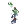
+ データを開く
- 基本情報
基本情報
| 登録情報 | データベース: PDB / ID: 3j6o | ||||||
|---|---|---|---|---|---|---|---|
| タイトル | Kinetic and Structural Analysis of Coxsackievirus B3 Receptor Interactions and Formation of the A-particle | ||||||
 要素 要素 | Coxsackie and adenovirus receptor | ||||||
 キーワード キーワード | CELL ADHESION / Coxsackievirus B3 / CVB3 / CAR | ||||||
| 機能・相同性 |  機能・相同性情報 機能・相同性情報AV node cell-bundle of His cell adhesion involved in cell communication / cell adhesive protein binding involved in AV node cell-bundle of His cell communication / homotypic cell-cell adhesion / AV node cell to bundle of His cell communication / epithelial structure maintenance / regulation of AV node cell action potential / gamma-delta T cell activation / transepithelial transport / germ cell migration / apicolateral plasma membrane ...AV node cell-bundle of His cell adhesion involved in cell communication / cell adhesive protein binding involved in AV node cell-bundle of His cell communication / homotypic cell-cell adhesion / AV node cell to bundle of His cell communication / epithelial structure maintenance / regulation of AV node cell action potential / gamma-delta T cell activation / transepithelial transport / germ cell migration / apicolateral plasma membrane / cell-cell junction organization / connexin binding / cardiac muscle cell development / heterophilic cell-cell adhesion via plasma membrane cell adhesion molecules / intercalated disc / bicellular tight junction / cell adhesion molecule binding / neutrophil chemotaxis / acrosomal vesicle / filopodium / mitochondrion organization / PDZ domain binding / Cell surface interactions at the vascular wall / adherens junction / neuromuscular junction / beta-catenin binding / Immunoregulatory interactions between a Lymphoid and a non-Lymphoid cell / cell-cell junction / integrin binding / cell junction / heart development / virus receptor activity / growth cone / cell body / actin cytoskeleton organization / basolateral plasma membrane / defense response to virus / neuron projection / membrane raft / signaling receptor binding / protein-containing complex / extracellular space / extracellular region / nucleoplasm / identical protein binding / plasma membrane / cytoplasm 類似検索 - 分子機能 | ||||||
| 生物種 |  Homo sapiens (ヒト) Homo sapiens (ヒト) | ||||||
| 手法 | 電子顕微鏡法 / 単粒子再構成法 / クライオ電子顕微鏡法 / 解像度: 9 Å | ||||||
 データ登録者 データ登録者 | Organtini, L.J. / Makhov, A.M. / Conway, J.F. / Hafenstein, S. / Carson, S.D. | ||||||
 引用 引用 |  ジャーナル: J Virol / 年: 2014 ジャーナル: J Virol / 年: 2014タイトル: Kinetic and structural analysis of coxsackievirus B3 receptor interactions and formation of the A-particle. 著者: Lindsey J Organtini / Alexander M Makhov / James F Conway / Susan Hafenstein / Steven D Carson /  要旨: The coxsackievirus and adenovirus receptor (CAR) has been identified as the cellular receptor for group B coxsackieviruses, including serotype 3 (CVB3). CAR mediates infection by binding to CVB3 and ...The coxsackievirus and adenovirus receptor (CAR) has been identified as the cellular receptor for group B coxsackieviruses, including serotype 3 (CVB3). CAR mediates infection by binding to CVB3 and catalyzing conformational changes in the virus that result in formation of the altered, noninfectious A-particle. Kinetic analyses show that the apparent first-order rate constant for the inactivation of CVB3 by soluble CAR (sCAR) at physiological temperatures varies nonlinearly with sCAR concentration. Cryo-electron microscopy (cryo-EM) reconstruction of the CVB3-CAR complex resulted in a 9.0-Å resolution map that was interpreted with the four available crystal structures of CAR, providing a consensus footprint for the receptor binding site. The analysis of the cryo-EM structure identifies important virus-receptor interactions that are conserved across picornavirus species. These conserved interactions map to variable antigenic sites or structurally conserved regions, suggesting a combination of evolutionary mechanisms for receptor site preservation. The CAR-catalyzed A-particle structure was solved to a 6.6-Å resolution and shows significant rearrangement of internal features and symmetric interactions with the RNA genome. IMPORTANCE: This report presents new information about receptor use by picornaviruses and highlights the importance of attaining at least an ∼9-Å resolution for the interpretation of cryo-EM ...IMPORTANCE: This report presents new information about receptor use by picornaviruses and highlights the importance of attaining at least an ∼9-Å resolution for the interpretation of cryo-EM complex maps. The analysis of receptor binding elucidates two complementary mechanisms for preservation of the low-affinity (initial) interaction of the receptor and defines the kinetics of receptor-catalyzed conformational change to the A-particle. | ||||||
| 履歴 |
|
- 構造の表示
構造の表示
| ムービー |
 ムービービューア ムービービューア |
|---|---|
| 構造ビューア | 分子:  Molmil Molmil Jmol/JSmol Jmol/JSmol |
- ダウンロードとリンク
- ダウンロード
| PDBx/mmCIF形式 |  3j6o.cif.gz 3j6o.cif.gz | 50.7 KB | 表示 |  PDBx/mmCIF形式 PDBx/mmCIF形式 |
|---|---|---|---|---|
| PDB形式 |  pdb3j6o.ent.gz pdb3j6o.ent.gz | 34.8 KB | 表示 |  PDB形式 PDB形式 |
| PDBx/mmJSON形式 |  3j6o.json.gz 3j6o.json.gz | ツリー表示 |  PDBx/mmJSON形式 PDBx/mmJSON形式 | |
| その他 |  その他のダウンロード その他のダウンロード |
-検証レポート
| 文書・要旨 |  3j6o_validation.pdf.gz 3j6o_validation.pdf.gz | 840.4 KB | 表示 |  wwPDB検証レポート wwPDB検証レポート |
|---|---|---|---|---|
| 文書・詳細版 |  3j6o_full_validation.pdf.gz 3j6o_full_validation.pdf.gz | 846.2 KB | 表示 | |
| XML形式データ |  3j6o_validation.xml.gz 3j6o_validation.xml.gz | 14.4 KB | 表示 | |
| CIF形式データ |  3j6o_validation.cif.gz 3j6o_validation.cif.gz | 19.1 KB | 表示 | |
| アーカイブディレクトリ |  https://data.pdbj.org/pub/pdb/validation_reports/j6/3j6o https://data.pdbj.org/pub/pdb/validation_reports/j6/3j6o ftp://data.pdbj.org/pub/pdb/validation_reports/j6/3j6o ftp://data.pdbj.org/pub/pdb/validation_reports/j6/3j6o | HTTPS FTP |
-関連構造データ
- リンク
リンク
- 集合体
集合体
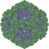
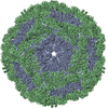
| 登録構造単位 | 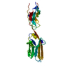
|
|---|---|
| 1 | x 60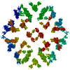
|
| 2 |
|
| 3 | x 5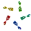
|
| 4 | x 6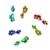
|
| 5 | 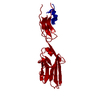
|
| 対称性 | 点対称性: (シェーンフリース記号: I (正20面体型対称)) |
- 要素
要素
| #1: タンパク質 | 分子量: 25046.229 Da / 分子数: 1 / 断片: SEE REMARK 999 / 由来タイプ: 組換発現 / 由来: (組換発現)  Homo sapiens (ヒト) / 発現宿主: Homo sapiens (ヒト) / 発現宿主:  |
|---|---|
| Has protein modification | Y |
| 配列の詳細 | THE HUMAN PROTEIN WAS IMAGED, BUT THE PROTEIN SEQUENCE THAT WAS FIT TO THE ELECTRON MICROSCOPY MAP ...THE HUMAN PROTEIN WAS IMAGED, BUT THE PROTEIN SEQUENCE THAT WAS FIT TO THE ELECTRON MICROSCOPY |
-実験情報
-実験
| 実験 | 手法: 電子顕微鏡法 |
|---|---|
| EM実験 | 試料の集合状態: PARTICLE / 3次元再構成法: 単粒子再構成法 |
- 試料調製
試料調製
| 構成要素 |
| ||||||||||||||||||||
|---|---|---|---|---|---|---|---|---|---|---|---|---|---|---|---|---|---|---|---|---|---|
| 分子量 | 値: 7 MDa / 実験値: YES | ||||||||||||||||||||
| ウイルスについての詳細 | 中空か: NO / エンベロープを持つか: NO / ホストのカテゴリ: VERTEBRATES / 単離: STRAIN / タイプ: VIRION | ||||||||||||||||||||
| 天然宿主 | 生物種: Homo sapiens | ||||||||||||||||||||
| 緩衝液 | 名称: 50 mM MES, 100 mM NaCl / pH: 6 / 詳細: 50 mM MES, 100 mM NaCl | ||||||||||||||||||||
| 試料 | 濃度: 0.1 mg/ml / 包埋: NO / シャドウイング: NO / 染色: NO / 凍結: YES | ||||||||||||||||||||
| 試料支持 | 詳細: glow-discharged holey carbon Quantifoil electron microscopy grids | ||||||||||||||||||||
| 急速凍結 | 装置: FEI VITROBOT MARK III / 凍結剤: OTHER / Temp: 95 K / 湿度: 95 % / 詳細: Plunged into ethane-propane (FEI VITROBOT MARK III) |
- 電子顕微鏡撮影
| 実験機器 |  モデル: Tecnai F20 / 画像提供: FEI Company |
|---|---|
| 顕微鏡 | モデル: FEI TECNAI F20 / 日付: 2012年8月1日 |
| 電子銃 | 電子線源:  FIELD EMISSION GUN / 加速電圧: 200 kV / 照射モード: SPOT SCAN FIELD EMISSION GUN / 加速電圧: 200 kV / 照射モード: SPOT SCAN |
| 電子レンズ | モード: BRIGHT FIELD / 倍率(公称値): 50000 X / 倍率(補正後): 50000 X / 最大 デフォーカス(公称値): 3660 nm / 最小 デフォーカス(公称値): 1980 nm / Cs: 2 mm / 非点収差: CTFFIND3 / カメラ長: 0 mm |
| 試料ホルダ | 試料ホルダーモデル: GATAN LIQUID NITROGEN / 傾斜角・最大: 0 ° / 傾斜角・最小: 0 ° |
| 撮影 | 電子線照射量: 15 e/Å2 / フィルム・検出器のモデル: KODAK SO-163 FILM |
| 画像スキャン | デジタル画像の数: 96 |
| 放射 | プロトコル: SINGLE WAVELENGTH / 単色(M)・ラウエ(L): M / 散乱光タイプ: x-ray |
| 放射波長 | 相対比: 1 |
- 解析
解析
| EMソフトウェア |
| ||||||||||||
|---|---|---|---|---|---|---|---|---|---|---|---|---|---|
| CTF補正 | 詳細: AUTO3DEM | ||||||||||||
| 対称性 | 点対称性: I (正20面体型対称) | ||||||||||||
| 3次元再構成 | 手法: Common Lines / 解像度: 9 Å / 解像度の算出法: FSC 0.5 CUT-OFF / 粒子像の数: 9302 / ピクセルサイズ(公称値): 1.25 Å / ピクセルサイズ(実測値): 1.25 Å 詳細: (Single particle details: Particles were selected using EMAN) (Single particle--Applied symmetry: I) 対称性のタイプ: POINT | ||||||||||||
| 原子モデル構築 | プロトコル: RIGID BODY FIT / 空間: REAL / Target criteria: correlation coefficient / 詳細: REFINEMENT PROTOCOL--rigid body | ||||||||||||
| 原子モデル構築 | PDB-ID: 3MJ7 PDB chain-ID: S / Accession code: 3MJ7 / Source name: PDB / タイプ: experimental model | ||||||||||||
| 精密化ステップ | サイクル: LAST
|
 ムービー
ムービー コントローラー



 PDBj
PDBj


