Movie
Movie Controller

[English] 日本語
 Yorodumi
- PDB-3j6m: Kinetic and Structural Analysis of Coxsackievirus B3 Receptor Int... -
+ Open data

- Basic information
| Entry | Database: PDB / ID: 3j6m | ||||||
|---|---|---|---|---|---|---|---|
| Title | Kinetic and Structural Analysis of Coxsackievirus B3 Receptor Interactions and Formation of the A-particle | ||||||
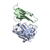
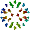
 Components Components | Coxsackievirus and adenovirus receptor | ||||||
 Keywords Keywords | CELL ADHESION / Coxsackievirus B3 / CVB3 / CAR | ||||||
| Function / homology |  Function and homology information Function and homology informationAV node cell-bundle of His cell adhesion involved in cell communication / cell adhesive protein binding involved in AV node cell-bundle of His cell communication / AV node cell to bundle of His cell communication / homotypic cell-cell adhesion / epithelial structure maintenance / regulation of AV node cell action potential / gamma-delta T cell activation / apicolateral plasma membrane / germ cell migration / connexin binding ...AV node cell-bundle of His cell adhesion involved in cell communication / cell adhesive protein binding involved in AV node cell-bundle of His cell communication / AV node cell to bundle of His cell communication / homotypic cell-cell adhesion / epithelial structure maintenance / regulation of AV node cell action potential / gamma-delta T cell activation / apicolateral plasma membrane / germ cell migration / connexin binding / transepithelial transport / cell-cell junction organization / cardiac muscle cell development / heterophilic cell-cell adhesion / intercalated disc / bicellular tight junction / cell adhesion molecule binding / neutrophil chemotaxis / acrosomal vesicle / Cell surface interactions at the vascular wall / mitochondrion organization / filopodium / adherens junction / PDZ domain binding / neuromuscular junction / beta-catenin binding / integrin binding / Immunoregulatory interactions between a Lymphoid and a non-Lymphoid cell / cell-cell junction / cell junction / heart development / cell body / growth cone / virus receptor activity / actin cytoskeleton organization / basolateral plasma membrane / defense response to virus / neuron projection / membrane raft / signaling receptor binding / protein-containing complex / extracellular space / extracellular region / nucleoplasm / identical protein binding / plasma membrane / cytoplasm Similarity search - Function | ||||||
| Biological species |  Homo sapiens (human) Homo sapiens (human) | ||||||
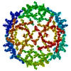
| Method | ELECTRON MICROSCOPY / single particle reconstruction / cryo EM / Resolution: 9 Å | ||||||
 Authors Authors | Organtini, L.J. / Makhov, A.M. / Conway, J.F. / Hafenstein, S. / Carson, S.D. | ||||||
 Citation Citation | Journal: J Virol / Year: 2014 Title: Kinetic and structural analysis of coxsackievirus B3 receptor interactions and formation of the A-particle. Authors: Lindsey J Organtini / Alexander M Makhov / James F Conway / Susan Hafenstein / Steven D Carson /  Abstract: The coxsackievirus and adenovirus receptor (CAR) has been identified as the cellular receptor for group B coxsackieviruses, including serotype 3 (CVB3). CAR mediates infection by binding to CVB3 and ...The coxsackievirus and adenovirus receptor (CAR) has been identified as the cellular receptor for group B coxsackieviruses, including serotype 3 (CVB3). CAR mediates infection by binding to CVB3 and catalyzing conformational changes in the virus that result in formation of the altered, noninfectious A-particle. Kinetic analyses show that the apparent first-order rate constant for the inactivation of CVB3 by soluble CAR (sCAR) at physiological temperatures varies nonlinearly with sCAR concentration. Cryo-electron microscopy (cryo-EM) reconstruction of the CVB3-CAR complex resulted in a 9.0-Å resolution map that was interpreted with the four available crystal structures of CAR, providing a consensus footprint for the receptor binding site. The analysis of the cryo-EM structure identifies important virus-receptor interactions that are conserved across picornavirus species. These conserved interactions map to variable antigenic sites or structurally conserved regions, suggesting a combination of evolutionary mechanisms for receptor site preservation. The CAR-catalyzed A-particle structure was solved to a 6.6-Å resolution and shows significant rearrangement of internal features and symmetric interactions with the RNA genome. IMPORTANCE: This report presents new information about receptor use by picornaviruses and highlights the importance of attaining at least an ∼9-Å resolution for the interpretation of cryo-EM ...IMPORTANCE: This report presents new information about receptor use by picornaviruses and highlights the importance of attaining at least an ∼9-Å resolution for the interpretation of cryo-EM complex maps. The analysis of receptor binding elucidates two complementary mechanisms for preservation of the low-affinity (initial) interaction of the receptor and defines the kinetics of receptor-catalyzed conformational change to the A-particle. | ||||||
| History |
|
- Structure visualization
| Movie |
Movie viewer |
|---|---|
| Structure viewer | Molecule: MolmilJmol/JSmol |
- Downloads & links
-Download
| PDBx/mmCIF format | 3j6m.cif.gz | 39.1 KB | Display | PDBx/mmCIF format |
|---|---|---|---|---|
| PDB format | pdb3j6m.ent.gz | 25.2 KB | Display | PDB format |
| PDBx/mmJSON format | 3j6m.json.gz | Tree view | PDBx/mmJSON format | |
| Others |  Other downloads Other downloads |
-Validation report
| Summary document | 3j6m_validation.pdf.gz | 851.8 KB | Display | wwPDB validaton report |
|---|---|---|---|---|
| Full document | 3j6m_full_validation.pdf.gz | 855.7 KB | Display | |
| Data in XML | 3j6m_validation.xml.gz | 13.4 KB | Display | |
| Data in CIF | 3j6m_validation.cif.gz | 17.7 KB | Display | |
| Arichive directory | https://data.pdbj.org/pub/pdb/validation_reports/j6/3j6mftp://data.pdbj.org/pub/pdb/validation_reports/j6/3j6m | HTTPS FTP |
-Related structure data
| Related structure data | 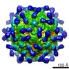 5927MC 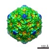 5928C 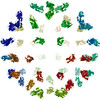 3j6lC 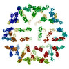 3j6nC 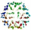 3j6oC M: map data used to model this data C: citing same article ( |
|---|---|
| Similar structure data |


-Links
 PDBj
PDBj


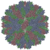
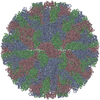
- Assembly
| Deposited unit | 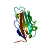
|
|---|---|
| 1 | x 60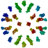
|
| 2 |
|
| 3 | x 5
|
| 4 | x 6
|
| 5 | 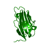
|
| Symmetry | Point symmetry: (Schoenflies symbol: I (icosahedral)) |
-Components
| #1: Protein | Mass: 13640.500 Da / Num. of mol.: 1 / Fragment: UNP residues 21-144 Source method: isolated from a genetically manipulated source Source: (gene. exp.) Homo sapiens (human) / Gene: CXADR, CAR / Production host:  |
|---|---|
| #2: Water | ChemComp-HOH /  Mass: 18.015 Da / Num. of mol.: 30 / Source method: isolated from a natural source / Formula: H2O Mass: 18.015 Da / Num. of mol.: 30 / Source method: isolated from a natural source / Formula: H2O |
| Has protein modification | Y |
-Experimental details
-Experiment
| Experiment | Method: ELECTRON MICROSCOPY |
|---|---|
| EM experiment | Aggregation state: PARTICLE / 3D reconstruction method: single particle reconstruction |
- Sample preparation
| Component |
| ||||||||||||||||||||
|---|---|---|---|---|---|---|---|---|---|---|---|---|---|---|---|---|---|---|---|---|---|
| Molecular weight | Value: 7 MDa / Experimental value: YES | ||||||||||||||||||||
| Details of virus | Empty: NO / Enveloped: NO / Host category: VERTEBRATES / Isolate: STRAIN / Type: VIRION | ||||||||||||||||||||
| Natural host | Organism: Homo sapiens | ||||||||||||||||||||
| Buffer solution | Name: 50 mM MES, 100 mM NaCl / pH: 6 / Details: 50 mM MES, 100 mM NaCl | ||||||||||||||||||||
| Specimen | Conc.: 0.1 mg/ml / Embedding applied: NO / Shadowing applied: NO / Staining applied: NO / Vitrification applied: YES | ||||||||||||||||||||
| Specimen support | Details: glow-discharged holey carbon Quantifoil electron microscopy grids | ||||||||||||||||||||
| Vitrification | Instrument: FEI VITROBOT MARK III / Cryogen name: OTHER / Temp: 95 K / Humidity: 95 % Details: Plunged into ethane-propane (FEI VITROBOT MARK III) |
- Electron microscopy imaging
| Experimental equipment |  Model: Tecnai F20 / Image courtesy: FEI Company |
|---|---|
| Microscopy | Model: FEI TECNAI F20 / Date: Aug 1, 2012 |
| Electron gun | Electron source:  FIELD EMISSION GUN / Accelerating voltage: 200 kV / Illumination mode: SPOT SCAN FIELD EMISSION GUN / Accelerating voltage: 200 kV / Illumination mode: SPOT SCAN |
| Electron lens | Mode: BRIGHT FIELD / Nominal magnification: 50000 X / Calibrated magnification: 50000 X / Nominal defocus max: 3660 nm / Nominal defocus min: 1980 nm / Cs: 2 mm / Astigmatism: CTFFIND3 / Camera length: 0 mm |
| Specimen holder | Specimen holder model: GATAN LIQUID NITROGEN / Tilt angle max: 0 ° / Tilt angle min: 0 ° |
| Image recording | Electron dose: 15 e/Å2 / Film or detector model: KODAK SO-163 FILM |
| Image scans | Num. digital images: 96 |
| Radiation | Protocol: SINGLE WAVELENGTH / Monochromatic (M) / Laue (L): M / Scattering type: x-ray |
| Radiation wavelength | Relative weight: 1 |
- Processing
| EM software |
| ||||||||||||
|---|---|---|---|---|---|---|---|---|---|---|---|---|---|
| CTF correction | Details: AUTO3DEM | ||||||||||||
| Symmetry | Point symmetry: I (icosahedral) | ||||||||||||
| 3D reconstruction | Method: Common Lines / Resolution: 9 Å / Resolution method: FSC 0.5 CUT-OFF / Num. of particles: 9302 / Nominal pixel size: 1.25 Å / Actual pixel size: 1.25 Å Details: (Single particle details: Particles were selected using EMAN) (Single particle--Applied symmetry: I) Symmetry type: POINT | ||||||||||||
| Atomic model building | Protocol: RIGID BODY FIT / Space: REAL / Target criteria: correlation coefficient / Details: REFINEMENT PROTOCOL--rigid body | ||||||||||||
| Atomic model building | PDB-ID: 1KAC Pdb chain-ID: B / Accession code: 1KAC / Source name: PDB / Type: experimental model | ||||||||||||
| Refinement step | Cycle: LAST
|